Overview of Small Intestine Histology and Function
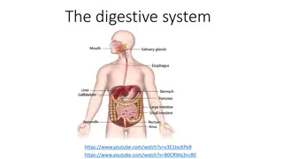
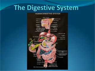
The small intestine is a key organ in the digestive system responsible for the digestion and absorption of nutrients. It is divided into the duodenum, jejunum, and ileum, each with specific functions and structures like plicae circulares, villi, microvilli, and crypts of Lieberkühn. The intestinal wall consists of the mucosa, lamina propria, and muscularis mucosa, housing essential cells like absorptive cells and goblet cells. Intestinal glands like the crypts of Lieberkühn contain stem cells, goblet cells, and Paneth cells contributing to digestion and protection against pathogens.

Download Presentation

Please find below an Image/Link to download the presentation.
The content on the website is provided AS IS for your information and personal use only. It may not be sold, licensed, or shared on other websites without obtaining consent from the author. Download presentation by click this link. If you encounter any issues during the download, it is possible that the publisher has removed the file from their server.
E N D
Presentation Transcript
Histology Digestive system By: Dr. Ammar Ismail
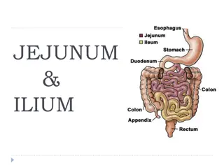
Small intestine The small intestine is divided into three regions : Duodenum , Jejunum , Ileum. the small intestine digests food material and absorbs end products of digestive process. To perform its digestive functions the first region of small intestine, the duodenum, receives enzymes and an alkaline buffer from the pancreas and bile from the liver. Additionally epithelial cells and glands of the mucosa contribute buffers and enzymes to facilitate digestion
Small intestine: The surface area of the intestinal lumen is enlarged by the formation of several important structures include: 1 Plicae circularis : are transverse folds of the sub mucosa and mucosa that form semicircular elevations . 2 Villi :Are epithelially covered, finger like protrusions of the lamina propria . Their numbers are greater in the duodenum than in the jejunum or ileum and their height decreases from duodenum to ileum . 3 Microvilli : modifications of the apical plasma membrane of the epithelial cells covering the intestinal villi , increase the surface area of the small intestine . 4 Crypt of lieberkuhn: Invagination of the epithelium into lamina propria between the villi form intestinal glands , which also increase the surface area of the small intestine .
Intestinal wall : 1 Intestinal mucosa: the mucosa of the small intestine is composed of the usual three layers; a simple columnar epithelium , lamina propria and the muscularis mucosa . Epithelium : The most numerous cells of epithelium are surface absorptive cells they are tall cells and its have brush border the principle function of these cells are terminal digestion and absorption . another cells are Goblet cells which are unicellular glands secrete mucin to protective layer lining the lumen . the number of these cells increase toward the ileum . The epithelium of small intestine is changed every 3 5 days and proliferate from regenerative cells in the crypt of liberkuhn
2 - Lamina propria : Its loose connective tissue contain intestinal gland (crept of lieberkuh ) which is simple tubular gland open into inter villar spaces .
Intestinal gland (crypt of lieberkuhn ): intestinal gland (crept of lieberkuhn) which is simple tubular gland open into inter villar spaces . It have three types cells : 1 Undifferentiated ( regenerative cells ) stem cells which are proliferate to provide epithelial cells . 2 Goblet cells: 3 Paneth cell :Pyramidal shaped cells occupy the bottom of crypts of lieberkuhn and manufacture the antibacterial agent lysozyme (peptidase ) the lamina propria also is rich in lymphoid tissue diffused or as lymph nodule which is increase in distal part of small intestine to form characteristic feature of Ileum (payer patch )
Muscularis mucosa of small intestine is composed of an inner circular layer and an outer longitudinal layer of smooth muscle . Sub mucosa: The sub mucosa of small intestine is composed of dense irregular fibro elastic connective tissue with rich lymphatic and vascular tissues also contain in the duodenum Brunner's glands Muscularis Externa : Its composed of an inner circular layer and an outer longitudinal smooth muscle layer , the muscularis externa is responsible for the peristaltic movement of small intestine . Serosa is present
Histological structure of the villus: The villus is a fold of small intestinal mucosa which consist of simple columnar epithelium ( absorptive ) with goblet cells and lamina propria form the core of villus . in this core there are three important structures which give the ability of absorption to the villi . 1 Capillaries loops under the epithelium to carry the amino acids and monosaccharide to the portal vein of liver . 2 Lacteal vessel : It s a blindly ending lymphatic channel in the center of villus to carry the absorptive fat to the large lymphatic vessels. 3 Smooth muscle fibers from the muscularis mucosa , the contraction of this fibers facilitate the passage of lymph in lacteal vessel
Large intestine: The wall of large intestine is composed of the same tunics of small intestine , although the large intestine is divided anatomically into several part but it have the same histological structure . Mucosa :The colon has no villi but has fold .The epithelium is simple columnar ( secretary) and large numbers of goblet cells (largest than small intestine ) and increase toward the distal part of large intestine . Lamina propria : Loss connective tissue it has intestinal gland crypt of lieberkhun which are similar in composition to those of small intestine except for the absence of Paneth cells . Muscularis mucosa and submucosa similar to that in small intestine . Muscularis externa :The arrangement of tunica muscularis is differ from that in small intestine especially the outer longitudinal layer is not continuous along the surface , it present as three longitudinal narrow ribbon which called taeniae coli . The serosa is present
Rectum and Anal canal : The rectum resemble the colon , the mucosa of anal canal has long fold and the crypt of lieberkuhn become short and absent gradually .The mucosa in the anus become stratified squamous Epithelium which transitional area between mucosa of intestine and skin . it contain sebaceous gland and circum anal gland and lamina propria in this area contain plexus of large veins . The mascularis externa in the anus consist of, internal and external anal sphinector muscle


:")
")