Understanding the Anatomy and Function of the Stomach
The stomach is a vital organ in the digestive system with functions like food storage, mixing with gastric secretions, and controlling chyme delivery to the small intestine. It has a J-shaped structure with various parts like the fundus, body, antrum, and pylorus. The lesser and greater curvatures, along with the cardiac and pyloric orifices, contribute to its unique shape and functionality. The pyloric sphincter regulates the outflow of gastric contents into the duodenum, crucial for digestion.

Download Presentation

Please find below an Image/Link to download the presentation.
The content on the website is provided AS IS for your information and personal use only. It may not be sold, licensed, or shared on other websites without obtaining consent from the author. Download presentation by click this link. If you encounter any issues during the download, it is possible that the publisher has removed the file from their server.
E N D
Presentation Transcript
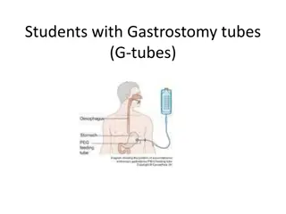
The Stomach Description The stomach is the dilated portion of the alimentary canal the three main functions: A. It stores food (in the adult it has a capacity of about 1500 ml). B. It mixes the food with gastric secretions to form a semifluid chyme. C. It controls the rate of delivery of the chyme to the small intestine . so that efficient digestion and absorption can take place. Position is situated in the upper part of the abdomen, extending from beneath the left costal margin region into the epigastric and umbilical regions. (Much of the stomach lies under cover of the lower ribs).
Shape of the stomach It is roughly J-shaped and has Two openings, the cardiac and pyloric orifices. Two curvatures, the greater and lesser curvatures. Two surfaces, an anterior and a posterior surface (Fig. 5.21). The stomach is relatively fixed at both ends but is very mobile in between. It tends to be high and transversely arranged in the short, obese person and elongated vertically in the tall, thin person. Its shape undergoes considerable variation in the same person and depends on the volume of its contents, the position of the body, and the phase of respiration.
Parts of the stomach 1. Fundus: is a dome-shaped and projects upward and to the left of the cardiac orifice. It is usually full of gas. 2. Body: This extends from the level of the cardiac orifice to the level of the incisura angularis, (constant notch in the lower part of the lesser curvature ). 3. Antrum: This extends from the incisura angularis to the pylorus (Fig. 5.21). 4. Pylorus: This is the most tubular part of the stomach. 5. The thick muscular wall is called the pyloric sphincter,
0ther parts: The lesser curvature forms the right border of the stomach and extends from the cardiac orifice to the pylorus. It is suspended from the liver by the lesser omentum. The greater curvature is much longer than the lesser curvature and extends from the left of the cardiac orifice, over the dome of the fundus, and along the left border of the stomach to the pylorus. The cardiac orifice is where the esophagus enters the stomach. Although no anatomic sphincter can be demonstrated here, a physiologic mechanism exists that prevents regurgitation of stomach contents into the esophagus. The pyloric orifice is formed by the pyloric canal, which is about 1 in. (2.5 cm) long.
Function of the Pyloric Sphincter Controls the outflow of gastric contents into the duodenum. 2. Receives motor fibers from the sympathetic system and inhibitory fibers from the vagi . 1.
Blood Supply of the stomach The arteries: are derived from the branches of the celiac artery. The left gastric artery arises from the celiac artery. It supplies the lower third of the esophagus and the upper right part of the stomach. The right gastric artery arises from the hepatic artery at the upper border of the pylorus and runs to the left along the lesser curvature. It supplies the lower right part of the stomach. The short gastric arteries arise from the splenic artery to supply the fundus. The left gastroepiploic artery arises from the splenic artery to supply the stomach along the upper part of the greater curvature. The right gastroepiploic artery arises from the gastroduodenal branch of the hepatic artery. supplies the stomach along the lower part of the greater curvature. 1. 2. 3. 4. 5.
Venous supply of the stomach The veins drain into the portal circulation (Fig. 5.22). The left and right gastric veins drain directly into the portal vein. The short gastric veins and the left gastroepiploic veins join the splenic vein. The right gastroepiploic vein joins the superior mesenteric vein
Nerve Supply of the stomach The nerve supply includes sympathetic fibers derived from the celiac plexus and parasympathetic fibers from the right and left vagus nerves . The anterior vagal trunk, which is formed in the thorax mainly from the left vagus nerve. The posterior vagal trunk, which is formed in the thorax mainly from the right vagus nerve . The sympathetic innervation of the stomach carries a proportion of pain-transmitting nerve fibers, whereas the parasympathetic vagal fibers are secretomotorto the gastric glands and motor to the muscular wall of the stomach.
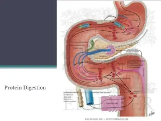
MICROSCOPIC ANATOMY OF THE STOMACH Most of the specialised cells of the stomach (parietal and chief Cells), also has numerous endocrine cells. Parietal cells These are in the body (acid-secreting portion) of the stomach and line the gastric crypts, They are responsible for the production of hydrogen ions to form hydrochloric acid. The hydrogen ions are actively secreted by the proton pump. Chief cells These lie principally proximally in the gastric crypts and produce pepsinogen.
3. Endocrine cells, which are critical to its function. In the gastric antrum, the mucosa contains G cells, which produce gastrin. Throughout the body of the stomach, enterochromaffin- like (ECL) cells are abundant and produce histamine, a key factor in driving gastric acid secretion.
Summary box The anatomy and physiology of the stomach The stomach acts as a reservoir for food and commences the process of digestion Gastric acid is produced by a proton pump in the parietal cells, which in turn is controlled by histamine acting on the H2-receptors. The histamine is produced by the endocrine gastric ECL cells in response to a number of factors, particularly gastrin and the vagus . Proton pump inhibitors abolish gastric acid production, whereas H2-receptor antagonists only markedly reduce it. The gastric mucous layer is essential to the integrity of the gastric mucosa
The investigation of gastric disorders Flexible endoscopy is the most commonly used and sensitive technique for investigating the stomach and duodenum. Great care needs to be exercised in performing endoscopy to avoid complications and missing important pathology. Axial imaging, particularly multi slice CT, is useful in the staging of gastric cancer, although it may be less sensitive in the detection of liver metastases than other modalities. Endoscopic ultrasound is the most sensitive technique in the evaluation of the T stage of gastric cancer and in the assessment of duodenal tumors. Laparoscopy is very sensitive in detecting peritoneal metastases, and laparoscopic ultrasound provides an accurate evaluation of lymph node and liver metastases 1. 2. 3. 4.
Alarm symptoms that indicate the need for upper endoscopy Age >55 years with new onset dyspepsia 1. 2. Unintentional weight loss 3. Persistent or recurrent vomiting 4. Progressive dysphagia 5. Recent onset odynophagia 6. Unexplained iron deficiency anemia or GI bleeding 7. Palpable abdominal mass or lymphadenopathy 8. Family history of upper gastrointestinal cancer