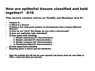
Introduction to Histology: Study of Cells, Tissues, and Organs
Histology, also known as microscopic anatomy, focuses on the study of cells, tissues, and organs through a microscope. It encompasses the examination of epithelial tissues, their special characteristics, functions, and embryological origins. Epithelial tissues play crucial roles in protection, absorption, secretion, and sensory functions. Derived from the three germ layers during embryonic development, epithelia exhibit distinct features such as cellularity, polarity, and regenerative capacities.

Download Presentation

Please find below an Image/Link to download the presentation.
The content on the website is provided AS IS for your information and personal use only. It may not be sold, licensed, or shared on other websites without obtaining consent from the author. Download presentation by click this link. If you encounter any issues during the download, it is possible that the publisher has removed the file from their server.
E N D
Presentation Transcript
Histology Study of cells, tissues and organs as seen with the help of microscope Study
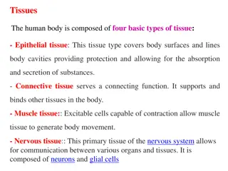
Histology Often called Microscopic Anatomy Greek word Histos= tissue Logia=science/study 4 basic type of tissues: Epithelial lining and covering Connective support Muscle movement Nervous control Cells work together in functionally related groups called tissues
Epithelial Tissue or Epithelium Consist of sheets of cells Covers a external surface of the body May line the internal cavities and the organs Forms most organs & glands
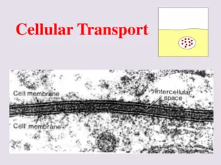
Special Characteristics of Epithelia Cellularity cells are in close contact with each other with little or no intercellular space between them Specialized contacts: Junctional Complexes may have junctions for both attachment and communication
Special Characteristics of Epithelia Polarity epithelial tissues always have an apical and basal surface Support by connective tissue at the basal surface, both the epithelial tissue and the connective tissue contribute to the basement membrane Avascular..No blood vessel, no lymphatics nutrients must diffuse
Special Characteristics of Epit Innervated Regeneration epithelial tissues have a high capacity for regeneration Invaginates and grows in the underlying CT..specializing as glands Apical surface shows modification: presence of cilia or microvilli Nuclear shape corresponds to the cell shape: oval in columnar,round in cuboidal or polyhedral and flat in squamous cells
Functions Functions of epithelium Protection Absorption, May secrete material and ion transport Filtration Forms slippery surfaces Function as sensory surfaces
Embryologically Epithelia are derived from all the 3 germ layers: Ectoderm Epithelium of skin Endoderm Epithelium of gut Mesoderm Epithelium of pericardial, peritoneal and pleural cavities
Basement Membrane All cells rest on it. Thin, non-cellular Separates epithelium from underlying connective tissue Easily seen with light microscope Made up of: Basal Lamina-Amorphous, product of epithelium Reticular Lamina-Reticular fibres, product of CT
Basement Membrane The epithelial cells lie on the reticular lamina (collagen CT) Reticular lamina is bound to another CT called areolarCT. Together this structure is called the "basement membrane
Basal Feature: The Basal Lamina Noncellular supporting sheet between the epithelium and the connective tissue deep to it Consists of proteins secreted by the epithelial cells Functions: Acts as a selective filter, determining which molecules from capillaries enter the epithelium Acts as scaffolding along which regenerating epithelial cells can migrate Basal lamina and reticular layers of the underlying connective tissue deep to it form the basement membrane
Intercellular Junctions OR Junctional Complexes Zonula occludens (Tight Junctions) Zonula adherens Macula adherens (Desmosome) and hemidesmosome Gap junction (Nexus)
Lateral Surface Features Factors holding epithelial cells together Adhesion proteins link plasma membranes of adjacent cells Contours of adjacent cell membranes Special cell junctions
Tight junctions (zona occludens) close off intercellular space Found at apical region of most epithelial types Some proteins in plasma membrane of adjacent cells are fused Prevent molecules from passing between cells of epithelial tissue ..Serves as a SELECTIVE BARRIER, giving it a sealing effect. Example- Intestine and urinary bladder
Adherens junctions (zonula adherens) anchoring junction Present just below the tight junctions Provides Rigidity to the apex of the cell. Presence of dense plaque like material on the cytoplasmic surface of the plasma membranes of the junction. Transmembrane linker proteins attach to actin microfilaments of the cytoskeleton and bind adjacent cells Along with tight junctions, form the tight junctional complex around apical lateral borders of epithelial tissues
Desmosomes (Macula Adherens) Hemidesmosomes Gap of 30nm Transmembrane Proteins Electron dense plaque Attachment to Intermediate Filaments FIRM ADHESION between cells Subjected to friction, Epidermis of skin.
Desmosomes Desmosomes two disc-like plaques connected across intercellular space Plaques of adjoining cells are joined by proteins called cadherins Proteins interdigitate into extracellular space Intermediate filaments insert into plaques from cytoplasmic side
Gap junctions (Nexus) passageway between two adjacent cells Let small molecules move directly between neighboring cells Cells are connected by hollow cylinders of protein Passage of inorganic ions Exchange of chemical messengers in cell recognition and differentiation.
Tight Junctions In the apical Band or belt Barrier device
Surface Modifications Stereocilia Long thick Microvilli, Non motile, may show branching, Increase surface area( Epididimis), helps perception of stimuli (Internal Ear) Cilia-long, hair like projections of plasma membrane Glycocalyx-rich in polysaccharides Concentrates ions prior to absorption Act as receptor sites for hormones and enzymes. Microvilli- minute finger like projections Increase absorptive surface
Microvilli and Cilia Nonmotile Contain Microfilaments Function-Absorption Intestinal epithelium, proximal convoluted tubules of the kidney Motile Contain 9+2 pattern of microtubules Driving the entangled particles, transport in one dcirection Examples: Respirastory tract,uterine tube and ependyma
Classifications & Naming of Epithelia According to the number of cell layers First name of tissue indicates number of layers Simple one layer of cells Stratified more than one layer of cells
Classification & Naming of Epithelia Last name of tissue describes shape of cells Squamous cells wider than tall (plate or scale like) Cuboidal cells are as wide as tall, as in cubes Columnar cells are taller than they are wide, like columns
Naming Epithelia Naming the epithelia includes both the layers (first) and the shape of the cells (second) i.e. stratified cuboidal epithelium The name may also include any accessory structures Goblet cells Cilia Keratin Special epithelial tissues (don t follow naming convention) Psuedostratified Transitional
Simple Squamous Epithelium Description single layer of flat cells with disc-shaped nuclei Special types Endothelium (inner covering) slick lining of hollow organs Mesothelium (middle covering) Lines peritoneal, pleural, and pericardial cavities Covers visceral organs of those cavities
Simple Squamous Epithelium Function Passage of materials by passive diffusion and filtration Secretes lubricating substances in serosae Location Renal corpuscles Alveoli of lungs Lining of heart, blood and lymphatic vessels Lining of ventral body cavity (serosae)
Simple Squamous Epithelium Simple squamous lining the walls of the capillary
Simple Cuboidal Epithelium Description single layer of cube-like cells with large, spherical central nuclei Function secretion and absorption Location kidney tubules, secretory portions of small glands, ovary & thyroid follicles
Simple Columnar Epithelium Description single layer of column-shaped (rectangular) cells with oval nuclei Some bear cilia at their apical surface May contain goblet cells Function Absorption; secretion of mucus, enzymes, and other substances Ciliated type propels mucus or reproductive cells by ciliary action
Simple Columnar Epithelium Location Non-ciliated form Lines digestive tract, gallbladder, ducts of some glands Ciliated form Lines small bronchi, uterine tubes, uterus
Pseudostratified Columnar Epithelium Description All cells originate at basement membrane Only tall cells reach the apical surface May contain goblet cells and bear cilia Nuclei lie at varying heights within cells Gives false impression of stratification Function secretion of mucus; propulsion of mucus by cilia
Pseudostratified Columnar Epithelium Locations Non-ciliated type Ducts of male reproductive tubes Ducts of large glands Ciliated variety Lines trachea and most of upper respiratory tract
Stratified Epithelia Contain two or more layers of cells Regenerate from below Major role is protection Are named according to the shape of cells at apical layer
Stratified Squamous Epithelium Description Many layers of cells squamous in shape Deeper layers of cells appear cuboidal or columnar Thickest epithelial tissue adapted for protection
Stratified Squamous Epithelium Specific types Keratinized contain the protective protein keratin Surface cells are dead and full of keratin Non-keratinized forms moist lining of body openings Function Protects underlying tissues in areas subject to abrasion Location Keratinized forms epidermis Non-keratinized forms lining of esophagus, mouth, and vagina
Transitional Epithelium Description Basal cells usually cuboidal or columnar Superficial cells dome- shaped or squamous Function stretches and permits distension of urinary bladder Location Lines ureters, urinary bladder and part of urethra
Introduction Histology There are (4) types of tissue: 1. Epithelial 2. Connective 3. Muscle 4. Nervous Similarities between tissue types: 1. All contain cells 2. Cells that make up tissues have similar functions
Epithelial Structure Apical Apical Basement Membrane
Basement Membrane The epithelial cells lie on the reticular lamina (collagen CT) Reticular lamina is bound to another CT called areolarCT. Together this structure is called the "basement membrane
Classification and Examples 1. SimpleEpithelium Single layer All cells anchored to basement membrane 2. Simple Squamous Kidney filtration 3. SimpleCuboidal Kidney tubules Filtration; secretion, absorption
Simple Epithelia 4. SimpleColumnar Tall, thin cells Absorptive cells (small intestine) Goblet Cells 5. Pseudostratified Ciliated Columnar Epithelium Pseudostratified ? Trachea Goblet Cells and Mucus
Stratified Epithelium 1. Characteristics 2+ layers 2. StratifiedSquamous Skin outer layer hardened by keratin 4 to 5 layers thick 3. StratifiedCuboidal Ducts of sweat glands This type + stratified columnar are rare!
MCQ Transitional epithelium is found in 1. Uterus 2. Ureter 3. Gall bladder 4. vagina


")
")
")
")


")