Histology of the Tongue: Structure and Function Overview
The tongue is covered by stratified squamous epithelium with various types of papillae on its dorsal surface, each serving specific functions such as taste perception and mechanical tasks. The filiform, fungiform, vallate, foliate, and circumvallate papillae are distinct in their appearance and distribution, contributing to taste sensation. Additionally, the intrinsic muscles of the tongue allow for its intricate movements, while taste buds located on gustatory papillae house different cell types for taste detection.

Uploaded on Sep 19, 2024 | 0 Views
Download Presentation

Please find below an Image/Link to download the presentation.
The content on the website is provided AS IS for your information and personal use only. It may not be sold, licensed, or shared on other websites without obtaining consent from the author. Download presentation by click this link. If you encounter any issues during the download, it is possible that the publisher has removed the file from their server.
E N D
Presentation Transcript
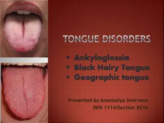
Histology of tongue The epithelium of dorsal surface of tongue is made up of stratified squamous epithelium which may be keratinized or non keratinized depending upon the species The epithelium of ventral side of tongue is made up of stratified squamous epithelium which is keratinized in all species The dorsal surface of the tongue consist of papillae which are grouped under gustatory and masticatory papillae
Filiform papillae It is ment for mechanical function and is lined with keratinized layer of SSE having central core of connective tissue extended up to tip in most animals In horses most of the papillae is formed by the epithelial prolongation with a small basal core of connective tissue Lenticular papillae They are found on torus linguae of ruminants and is made up of SSE which is keratinized.The central core of connective tissue presents secondary branching. Conical papillae They are similar to the conical papillae of the cheek but have less degree of keratinization
Fungiform papillae They are mushroom like papillae and are distributed over the dorsal surface and lined with SSE which is non keratinized It presents taste buds and the central core of connective tissue may show secondary branching with numerous nerve fibers Vallate papillae They are normally not extended over the dorsum linguae. They are surrounded by circular trench or moat. The surface epithelium consist of SSE which is non keratinized. Numerous taste buds are situated near the moat.They are classified under gustatory papillae.
FILIFORMAND FUNGIFORM PAPILLAE
CIRCUMVALLATE PAPILLAE
Foliate papillae They are situated towards palatoglossal arch.They are leaf like arranged in several rows and the gap between two row is known as gustatory furrow.They are lined with SSE of non keratinized variety. The taste buds are distributed along the epithelium of the sides facing the gustatory furrow Muscles of tongue As per the orientation the intrinsic muscles are of three variety and classified as longitudinal, vertical and transverse which are responsible for high mobility of tongue Taste buds They are oval or elliptical in shape and are distributed along the epithelium of gustatory papillae. The free end consist of a taste pore.Their are three types of cells present mainly taste hair cells,sustentacular cells and basal cells.
Taste hair cells are elongated and faces the taste pore and consist of fine hair like cytoplasmic processes The basal end is blunt and are associated with sensory nerve fibers which extend along the central core of papillae. They are responsible for carrying the sensation of taste The supporting cells are responsible for protection of taste hair cells. The basal cells are oval or polyhedral and are situated at the base of taste buds and are supposed to be progenitor of other two cell types
Lyssa It is a cord like structure formed by congregation of connective tissue, skeletal muscles and fat and the term is used in case of dog In cat similar structure is found and is predominantly filled with adipose tissue In case of pig a cord like structure is present at the sub epithelial layer of dorsum linguae and mainly consist of hyaline cartilage Teeth It consist of enamel,dentine,cementum and pulp Enamel is derived from ectoderm and the cells involved in its formation are called ameloblast cells Dentine is formed by the cells of mesodermal origin which are known as odontoblast
The odontoblast are arranged along the inner margin of dentine at the junction of pulp cavity. During mineralization some of the areas of dentine remains unmineralized throughout life and these areas constitute the interglobular spaces of owen The cementum is formed by mesodermal cells identified as cementoblast which is very much similar to osteoblast and as such it appears like that of osseous tissue. It is covered from outside by thick layer of connective tissue known as periodontal membrane consisting mainly of collagen fibers Pulp is the central cavity of the tooth surrounded by dentine and is formed by loosely packed mesenchymal cells along with nerve filaments and blood vessels