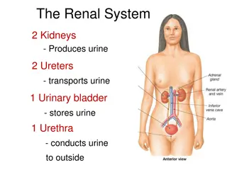
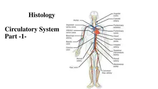
Histology of the Renal System for OSPE Exam Preparation

This comprehensive OSPE file for renal block covers key structures of the kidney including the cortex, juxtaglomerular apparatus, renal corpuscle, and kidney medulla. It provides detailed identification of structures, labeling of components, and differentiation between tubules. Helpful images and descriptions aid in understanding the histology of the renal system.

Download Presentation

Please find below an Image/Link to download the presentation.
The content on the website is provided AS IS for your information and personal use only. It may not be sold, licensed, or shared on other websites without obtaining consent from the author. Download presentation by click this link. If you encounter any issues during the download, it is possible that the publisher has removed the file from their server.
E N D
Presentation Transcript
OSPE file (Renal block) Red: questions. Dark red: very important. Black: complete answers. Gray: notes|extra.
You should know before the exam: The diagrams in these slides are going to be the same in the exam however, it may not be coloured. You have to mention the full name always and don t use shortcuts you could lose marks because of that. The Arrows in the diagrams are very important . So please study them well. Practical histology (OSPE) team 437 | Renal block
Cortex of the Kidney Q1: Identify the structure? Cortex of the Kidney Q2: Identify labeling: 1. Glomerulus of renal corpuscle. 2. Urinary space (or capsular space). 3. Parietal layer of Bowman s capsule. 4. Proximal convoluted tubules with brush border. 5. Distal convoluted tubules. 6. Juxta-glomerular apparatus. Q3: How to differentiate between the proximal and distal convoluted tubules? PCV: Has an ill defined lumen because it is filled with brush border (microvilli) DCV: Has a well demarcated and clear lumen. Practical histology (OSPE) team 437 | Renal block
Juxtaglomerular apparatus Q1: Identify the structure? Juxtaglomerular apparatus Q2: What are the components(cells) that form this structure? Juxtaglomerular cells of afferent glomerular arteriole (they secrete renin) afferent arteriole The macula densa of distal tubule (tall columnar cells) Distal tubule The extraglomerular mesangial cells Practical histology (OSPE) team 437 | Renal block
Renal Corpuscle Q1: Identify the structure? Renal Corpuscle Q2: What are the components that form this structure? Glomerulus which contains fenestrated capillaries "without diaphragm Bowman s capsule: Parietal layer, urinary space, and podocytes (visceral layer) Mesangial cells: Intra-glomerular cells DCT Q3: What are the cells that form this structure? Mesangial cells Glomerular endothelial cells Podocytes (epithelial cells) PCT DCT PCT DCT Practical histology (OSPE) team 437 | Renal block
Kidney (Medulla) Q1: Identify the structure? Kidney (Medulla) Q2: What is the features of the structure? Contains tubular structures: loop of Henle: simple squamous epithelium collecting tubules and ducts: simple cuboidal epithelium Papillary ducts (ducts of Bellini): Simple Columnar epithelium Practical histology (OSPE) team 437 | Renal block
Blood renal Barrier Q1: Identify the structure? Blood renal Barrier (Glomerular Filtration Barrier) Q2: What are the components that form this structure? 1. Endothelial wall of the glomerular capillaries. 2. The glomerular basal lamina (inner and outer laminae rarae and middle lamina densa). 3. Visceral layer of Bowman s capsule (podocytes). Practical histology (OSPE) team 437 | Renal block
Ureter Q1: Identify the structure? Ureter Q2: What is the type of epithelium in this structure? Transitional epithelium Q3: What is the features of the structure? Transitional epithelium Inner longitudinal layer Outer circular Blood vessels Adventitia Practical histology (OSPE) team 437 | Renal block
Urinary bladder Q1: Identify the structure? Urinary bladder Q2: What is the type of epithelium in this structure? Transitional epithelium Q3: What is the features of the structure? Transitional epithelium Lamina propria Connective tissue Mesothelium *It is a continuation of the lower third of the ureter, has the same structure except that it has serosa. Practical histology (OSPE) team 437 | Renal block
Layers Mucosa Muscularis Adventitia Upper 2/3: Inner Longitudinal Outer Circular Transitional Epithelium Lamina Propria Ureter NO Serosa Lower 1/3: Inner Longitudinal Middle Circular Outer Longitudinal 3 layers of smooth muscle coat Inner Longitudinal Middle Circular Outer Longitudinal *The name of this layer is urinary bladder is serosa or adventitia Serosa Transitional Epithelium Lamina Propria Urinary bladder Practical histology (OSPE) team 437 | Renal block
Team leaders : Rawan Mohammad Alharbi Khalid Fayez Alshehri Twitter.com/Histology437 HistologyTeam437@gmail.com
")


")