Understanding Epithelial Tissue in General Histology

Epithelial tissue is a crucial type of tissue in the human body, performing functions like protection, absorption, secretion, and sensation detection. This tissue is composed of cells with minimal extracellular matrix and plays a vital role in various bodily processes. Learn about the classification and functions of epithelial tissue, as well as its cellular structure and importance in histology.

Download Presentation

Please find below an Image/Link to download the presentation.
The content on the website is provided AS IS for your information and personal use only. It may not be sold, licensed, or shared on other websites without obtaining consent from the author. Download presentation by click this link. If you encounter any issues during the download, it is possible that the publisher has removed the file from their server.
E N D
Presentation Transcript
SECOND STAGE\GENERAL HISTOLOGY LEC2/ EPITHELIAL TISSUE . .
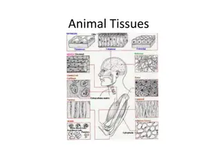
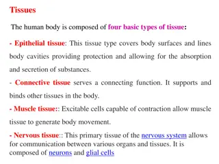
EPITHELIAL TISSUE Tissues are: Collections of specialized cells and cell products organized to perform a limited number of functions Histology = study of tissues. There are four main types of tissue in the human body 1-epithelial tissue 2-connective tissue 3-nervous tissue 4-muscular tissue
All these tissue composed from cells and extracellular matrix .epithelial tissue are composed of cells and very little extracellular matrix. these cells have strong adhesion and form cellular sheets that covered the surface of the body and line its cavities .cells of epithelial tissue supported by basement membrane of variable thickness . Basement membrane separate epithelia from underlying supporting tissue and never penetrated by blood vessels. thus epithelia depending on the diffusion of oxygen and metabolisms from adjacent supporting tissue .
THE FUNCTION OF EPITHELIAL TISSUE : Protection for the underlying tissues from radiation, desiccation, toxins, and physical trauma. e.g. skin 1. Absorption of substances in the digestive tract lining with distinct modifications. e.g. intestine 2. Regulation and excretion of chemicals between the underlying tissues and the body cavity. .e.g. goblet cell 3. The secretion of hormones into the blood vascular system. The secretion of sweat, mucus, enzymes, and other products that are delivered by ducts come from the glandular epithelium eg. Sweet glands 4. The detection of sensation. .e.g. taste buds 5.
CLASSIFICATION OF EPITHELIAL TISSUE -Classify epithelial tissue by cell shape and layers According to the number of cells : Simple epithelium Stratified epithelium According to the shape of cells : Squamous epithelium Cuboidal epithelium Columnar epithelium
Simple epithelium composed of single layer . and it defined as surface epithelia consisting from one layer of cell . and it range in shape from extremely flattened to tall columnar depending on its function
1. Simple squamous is flattened irregularly shaped cells forming continues surface that referred to as pavement membrane .this cells supporting by underlying layer basement membrane .simple squamous found lining surface such as in pleural ,pericardial ,peritoneal.
2- Simple Cuboidal epi. It is intermediate form between simple squamous and simple columnar the nucleus round and located in the center of cell .this type usually lines small ducts and tubules which have excretory , secretory or absorption function .e.g. small ducts of the kidney ,salivary gland and pancreas
3- Simple columnar epi.: is similar to cuboidal except that cells are taller and appear columnar in section . the height of cells may vary from tall to low depending on site and degree of functional activity . the nuclei are elongated and may be located towards the base . simple columnar found on highly absorption surface e.g. small intestine although secretory surface such as stomach . in this type may cell have cilia on the majority of the cells . among this ciliated cells are scattered non ciliated cells which have secretory function .simple columnar ciliated cell is not common in human except in the female reproductive tract. E.g. fallopian tube
4-Pseudostratified columnar ciliated epi .:in this type the cell rest on basement membrane and the nuclei of these cells are disposed at different levels thus creating the illusion pf cellular stratification this type found in larger airways of respiratory system e.g. bronchus.
Stratified epithelium : composed more than layer of cells this type have protective function and the degree and nature of the stratification are related to the kind of physical stress to which the surface is exposed . the classification of stratified epi . is based on the shape and structure of the surface cells since cells of the basal layer are usually cuboidal in shape .
1. Stratified squamous epithelium: consist on variable number of cells layers which exhibit transition from a cuboidal cell in basal to a flattened surface cells . the cells in the basal are divided continuously to replace the damage cells .this type found in oral cavity . esophagus ,uterine ,cervix and vagina .
2-Stratified squamous keratinizing epithelium: specialized form found in the epidermis in the surface of the skin .it is tough nonliving surface layer consisting of protein keratin
3-Stratified cuboidal epithelium: is a thin stratified epithelium which usually consist of only two or three layers of cuboidal or low columnar cells . this type of epi. Is usually confined to the the lining of the larger excretory ducts of exocrine glands such as salivary glands .
4- transitional epithelium: is a form of stratified epi . almost exclusively in the urinary tract. it is named because it has some features which intermediate (transitional ) between stratified cuboidal and stratified squamous . in the relaxed (contacted ) state transitional epi . appears to be about 4to 5 cells layer thick . the basal cells are roughly cuboidal , the intermediate cells are polygonal and the surface cells are large and rounded and may contain two nuclei . in the stretched state transitional epi. Appears only two or three cells thick . and the intermediate and surface layers are flattened.
SPECIALIZATIONS OF THE APICAL SURFACE Structures that Protruding from the apical surfaces of epithelial cells ,& Increase the cell surface area, or move substance from epithelia.
A. Microvilli: - Finger-like extensions. - Seen in absorptive cells; small intestine and proximal renal tubule. - Contain a core bundle of actin filaments. LM; Brush border.
B. Steriocilia Long, non-motile extensions. Long and branched microvilli. Seen in Epididymis and ducts deferens. Movement of molecules in and out of cells.
C. Cilia: is an organelle has shape of a slender protuberance that projects from the much larger cell body. There are two major types of cilia: motile and non-motile cilia. Non-motile cilia are also called primary cilia which serve as sensory organelles.
D.Flagellum: Only in spermatozoa, longer and one per cell.
MOST CELLS ARE STATIONARY MOST CELLS ARE STATIONARY- - HELD IN PLACE BY CELL JUNCTIONS: BY CELL JUNCTIONS: HELD IN PLACE 1. Tight junctions-zonula occludens Most apical junction form fluid tight seals in epithelium (close of intercellular space) that Line surfaces of organs and body cavities .Tight junctions prevent contents of organs from leaking into the blood & surrounding tissue . found in form of belt completely encircling the cell as zonula.
2-Adhering junction (zonula adherens) Encircle the cell . Made of plaque, to which attached actin filament that belong to terminal web . Cross the space between membranes and connect with proteins of an adjacent cell . Helps cell resist separation.
3-Desmosome- Desmosomes are specialized and highly ordered membrane domains that mediate cell-cell contact and strong adhesion. Adhesive interactions at the desmosome are coupled to the intermediate filament cytoskeleton. Spot welds also composed of plaque. Glycoproteins that help cells attach to each other.
4- Hemidesmosomes multiprotein complexes that facilitate the stable adhesion of basal epithelial cells to the underlying basement membrane. The mechanical stability of hemidesmosomes relies on multiple interactions of a few protein components that form a membrane-embedded tightly-ordered complex. Helps connect cells to the basement membrane. Helps anchor one kind of tissue to another in the body.
5- Gap junction Communicating Junctions- Gap junctions. Allow cells in a tissue to communicate. Allow chemical or electrical signals to pass between cells. Could found anywhere along the lateral surface. Found in all mammalian tissue except skeletal muscle. Individual unit called connexin, which made by 6 gap junction protein connexins.


 Encircle the cell .")