Understanding the Anatomy of the Small Intestine: Duodenum, Jejunum, and Ilium
 undefined


S
m
a
l
l
I
n
t
e
s
t
i
n
e
P
R
E
S
E
N
T
E
D
B
Y
M
S
C
.
D
R
.
R
E
H
A
M
S
A
A
D
K
A
D
H
U
M


S
m
a
l
l
I
n
t
e
s
t
i
n
e
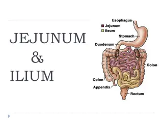
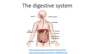
The small intestine is the longest part of the alimentary canal and extends from the pylorus of the stomach to the
ileocecal junction .
It is divided into three parts: the duodenum, the jejunum, and Ilium .
A. Duodenum ( fixed part )
Location and Description
1.The duodenum is a C-shaped tube, 2. about 10 in. (25 cm)long, which joins the stomach to the jejunum.
3.The duodenum curves around the head of the pancreas 4. The first inch (2.5 cm) of the duodenum resembles
the stomach in that it is covered on its anterior and posterior surfaces with peritoneum and has the lesser
omentum attached to its upper border and the greater omentum attached to its lower border; the lesser sac lies
behind this short segment. The remainder of the duodenum is
retroperitoneal
, being only
partially covered by
peritoneum.
P
a
r
t
s
o
f
t
h
e
D
u
o
d
e
n
u
m
The duodenum is situated in the epigastric and umbilical regions and it is divided into four parts.
First Part of the Duodenum
begins at the pylorus and runs upward and backward on the transpyloric plane at the
level of
the 1st lumbar vertebra
The relations of this part are as follows:
■■
Anteriorly
: The quadrate lobe of the liver and the gallbladder
■■
Posteriorly
: The lesser sac (first inch only), the gastroduodenal artery, the bile duct and the portal vein, and
the inferior vena cava
■■
Superiorly:
The entrance into the lesser sac (the epiploic foramen)
■■
Inferiorly
: The head of the pancreas
S
e
c
o
n
d
P
a
r
t
o
f
t
h
e
D
u
o
d
e
n
u
m
The second part of the duodenum runs vertically downward in front of the hilum of the right kidney on the
right side of the 2nd and 3rd lumbar vertebrae. About halfway down its medial border, the bile duct and the
main pancreatic duct pierce the duodenal wall. They unite to form the ampulla that opens on the summit of
the major duodenal papilla .The accessory pancreatic duct, if present, opens into the duodenum a little higher
up on the minor duodenal papilla .
The relations of this part are as follows:
■■
Anteriorly: The fundus of the gallbladder and the right lobe of the liver, the transverse colon, and the coils
of the
small intestine
■■
Posteriorly: The hilum of the right kidney and the right ureter
■■
Laterally: The ascending colon, the right colic flexure, and the right lobe of the liver
■■
Medially: The head of the pancreas, the bile duct, and the main pancreatic duct
T
h
i
r
d
P
a
r
t
o
f
t
h
e
D
u
o
d
e
n
u
m
The third part of the duodenum runs horizontally to the left on the subcostal plane, passing in front
of the vertebral column and following the lower margin of the head of the pancreas .
The relations of this part are as follows:
■■
Anteriorly:
The root of the mesentery of the small intestine, the superior mesenteric vessels
contained within it, and coils of jejunum
■■
Posteriorly
: The right ureter, the right psoas muscle, the inferior vena cava, and the aorta
■■
Superiorly
: The head of the pancreas
■■
Inferiorly:
Coils of jejunum
F
o
u
r
t
h
P
a
r
t
o
f
t
h
e
D
u
o
d
e
n
u
m
The fourth part of the duodenum runs upward and to the left to the duodenojejunal
flexure .The flexure is held in position by a peritoneal fold, the ligament of Treitz, which is
attached to the right crus of the diaphragm
The relations of this part are as follows:
Anteriorly: The beginning of the root of the mesentery and coils of jejunum .
Posteriorly: The left margin of the aorta and the medial border of the left psoas muscle
M
u
c
o
u
s
M
e
m
b
r
a
n
e
a
n
d
D
u
o
d
e
n
a
l
P
a
p
i
l
l
a
e
The mucous membrane of the duodenum is thick. In the first part of the duodenum, it is
smooth. In the remainder of the duodenum, it is thrown into numerous circular folds called the
plicae circulares. At the site where the bile duct and the main pancreatic duct pierce the medial
wall of the second part is a small, rounded elevation called the major duodenal papilla .
The accessory pancreatic duct, if present, opens into the duodenum on a smaller papilla about
0.75 in. (1.9 cm) above the major duodenal papilla.
B
l
o
o
d
S
u
p
p
l
y
&
V
e
i
n
s
Blood Supply
Arteries The upper half is supplied by the superior pancreaticoduodenal artery, a branch of the
gastroduodenal artery .The lower half is supplied by the inferior pancreaticoduodenal artery, a branch of the
superior mesenteric artery.
Veins
The superior pancreaticoduodenal vein drains into the portal vein; the inferior vein joins the superior
mesenteric vein .
Lymph Drainage
The lymph vessels follow the arteries and drain upward via
pancreaticoduodenal nodes to the gastroduodenal nodes and then to the celiac
nodes and downward via pancreaticoduodenal nodes to the superior mesenteric
nodes around the origin of the superior mesenteric artery.
Nerve Supply
The nerves are derived from sympathetic and parasympathetic (vagus) nerves
from the celiac and superior mesenteric plexuses.
J
e
j
u
n
u
m
a
n
d
I
l
e
u
m
(
m
o
b
i
l
e
p
o
r
t
i
o
n
)
Location and Description
The jejunum and ileum measure about 20 ft (6 m) long; the upper two fifths of this length make
up the jejunum. Each has distinctive features, but there is a gradual change from one to the
other. The jejunum begins at the duodenojejunal flexure, and the ileum ends at the ileocecal
junction. The coils of jejunum and ileum are freely mobile and are attached to the posterior
abdominal wall by a fan-shaped fold of peritoneum known as the mesentery of the small
intestine .
◦
The End
The small intestine is a vital part of the digestive system, comprising the duodenum, jejunum, and ileum. The duodenum, the fixed part, has specific anatomical features and relations with adjacent structures. Understanding the different parts of the duodenum, including their location and relationships with surrounding organs, is crucial for comprehension of its functions in digestion.

Download Presentation

Please find below an Image/Link to download the presentation.
The content on the website is provided AS IS for your information and personal use only. It may not be sold, licensed, or shared on other websites without obtaining consent from the author. Download presentation by click this link. If you encounter any issues during the download, it is possible that the publisher has removed the file from their server.
E N D
Presentation Transcript
Small Intestine PRESENTED BY MSC. DR. REHAM SAAD KADHUM
Small Intestine Small Intestine The small intestine is the longest part of the alimentary canal and extends from the pylorus of the stomach to the ileocecal junction . It is divided into three parts: the duodenum, the jejunum, and Ilium . A. Duodenum ( fixed part ) Location and Description 1.The duodenum is a C-shaped tube, 2. about 10 in. (25 cm)long, which joins the stomach to the jejunum. 3.The duodenum curves around the head of the pancreas 4. The first inch (2.5 cm) of the duodenum resembles the stomach in that it is covered on its anterior and posterior surfaces with peritoneum and has the lesser omentum attached to its upper border and the greater omentum attached to its lower border; the lesser sac lies behind this short segment. The remainder of the duodenum is retroperitoneal, being only partially covered by peritoneum.
Parts of the Duodenum The duodenum is situated in the epigastric and umbilical regions and it is divided into four parts. First Part of the Duodenum begins at the pylorus and runs upward and backward on the transpyloric plane at the level of the 1st lumbar vertebra The relations of this part are as follows: Anteriorly: The quadrate lobe of the liver and the gallbladder Posteriorly: The lesser sac (first inch only), the gastroduodenal artery, the bile duct and the portal vein, and the inferior vena cava Superiorly: The entrance into the lesser sac (the epiploic foramen) Inferiorly: The head of the pancreas
Second Part of the Duodenum The second part of the duodenum runs vertically downward in front of the hilum of the right kidney on the right side of the 2nd and 3rd lumbar vertebrae. About halfway down its medial border, the bile duct and the main pancreatic duct pierce the duodenal wall. They unite to form the ampulla that opens on the summit of the major duodenal papilla .The accessory pancreatic duct, if present, opens into the duodenum a little higher up on the minor duodenal papilla . The relations of this part are as follows: Anteriorly: The fundus of the gallbladder and the right lobe of the liver, the transverse colon, and the coils of the small intestine Posteriorly: The hilum of the right kidney and the right ureter Laterally: The ascending colon, the right colic flexure, and the right lobe of the liver Medially: The head of the pancreas, the bile duct, and the main pancreatic duct
Third Part of the Duodenum The third part of the duodenum runs horizontally to the left on the subcostal plane, passing in front of the vertebral column and following the lower margin of the head of the pancreas . The relations of this part are as follows: Anteriorly: The root of the mesentery of the small intestine, the superior mesenteric vessels contained within it, and coils of jejunum Posteriorly: The right ureter, the right psoas muscle, the inferior vena cava, and the aorta Superiorly: The head of the pancreas Inferiorly: Coils of jejunum
Fourth Part of the Duodenum The fourth part of the duodenum runs upward and to the left to the duodenojejunal flexure .The flexure is held in position by a peritoneal fold, the ligament of Treitz, which is attached to the right crus of the diaphragm The relations of this part are as follows: Anteriorly: The beginning of the root of the mesentery and coils of jejunum . Posteriorly: The left margin of the aorta and the medial border of the left psoas muscle
Mucous Membrane and Duodenal Papillae The mucous membrane of the duodenum is thick. In the first part of the duodenum, it is smooth. In the remainder of the duodenum, it is thrown into numerous circular folds called the plicae circulares. At the site where the bile duct and the main pancreatic duct pierce the medial wall of the second part is a small, rounded elevation called the major duodenal papilla . The accessory pancreatic duct, if present, opens into the duodenum on a smaller papilla about 0.75 in. (1.9 cm) above the major duodenal papilla.
Blood Supply & Veins Blood Supply Arteries The upper half is supplied by the superior pancreaticoduodenal artery, a branch of the gastroduodenal artery .The lower half is supplied by the inferior pancreaticoduodenal artery, a branch of the superior mesenteric artery. Veins The superior pancreaticoduodenal vein drains into the portal vein; the inferior vein joins the superior mesenteric vein .
Lymph Drainage The lymph vessels follow the arteries and drain upward via pancreaticoduodenal nodes to the gastroduodenal nodes and then to the celiac nodes and downward via pancreaticoduodenal nodes to the superior mesenteric nodes around the origin of the superior mesenteric artery. Nerve Supply The nerves are derived from sympathetic and parasympathetic (vagus) nerves from the celiac and superior mesenteric plexuses.
Jejunum and Ileum ( mobile portion ) Location and Description The jejunum and ileum measure about 20 ft (6 m) long; the upper two fifths of this length make up the jejunum. Each has distinctive features, but there is a gradual change from one to the other. The jejunum begins at the duodenojejunal flexure, and the ileum ends at the ileocecal junction. The coils of jejunum and ileum are freely mobile and are attached to the posterior abdominal wall by a fan-shaped fold of peritoneum known as the mesentery of the small intestine .


")
")
")