Understanding the Anatomy of the Large Intestine


Objectives
At the end of the lecture, students should be able to:
List the different
parts
of
large intestine
.
List the
characteristic
features
of
colon
.
Describe the anatomy of
different parts
of
large intestine
regarding:
the
surface anatomy
,
peritoneal covering
,
relations
,
arterial
&
nerve
supply
.
o
(1,2,3,4,5) are found in the
abdomen
o
(6,7) are found in the
pelvis
o
(8) is found in the
perineum
o
NOT found in rectum & anal canal ONLY colon
o
Taeniae coli
:
Three
longitudinal muscle bands
o
Sacculations (
Haustra
)
=
تكيّسات
:
Because the Taeniae coli are
shorter
than large intestine
o
Epiploic Appendices
زوائد=
:
Short
peritoneal folds
filled with fat
(Yellowish color)
P
a
r
t
s
o
f
t
h
e
L
a
r
g
e
I
n
t
e
s
t
i
n
e
C
h
a
r
a
c
t
e
r
i
s
t
i
c
s
o
f
C
o
l
o
n
1- The peritoneum covers the anterior and posterior surfaces.
2- The peritoneum only covers the anterior surface
3- First 2 inches WITHOUT mesentery
P
e
r
i
t
o
n
e
a
l
C
o
v
e
r
i
n
g
P
a
r
t
s
o
f
t
h
e
L
a
r
g
e
I
n
t
e
s
t
i
n
e
R
e
l
a
t
i
o
n
*Transverse colon
full
Stomach
will be
Anterior
*Transverse colon
empty
Stomach
will be
Superior
C
o
l
i
c
f
l
e
x
u
r
e
s
&
A
p
p
e
n
d
i
x
(REMEMBER! The position of base not change)
*In case of
appendicitis
this area will have
rebound tenderness
(It refers to
pain
upon
removal
of
pressure
rather
than
application of
pressure
to
the abdomen
)
R
e
c
t
u
m
- Upper 1/3 of rectum is covered by peritoneum in front & side
- Middle 1/3 of rectum is covered by peritoneum in front ONLY
- Lower 1/3 of rectum & anal canal has no peritoneum cover
R
e
l
a
t
i
o
n
b
e
t
w
e
e
n
e
m
b
r
y
o
l
o
g
i
c
a
l
o
r
i
g
i
n
o
f
G
I
T
&
S
u
p
p
l
y
*It continues as the
superior rectal artery
in the root of the
sigmoid mesocolon (pelvic region).
1. The taeniae coli found in which of the following structure?
A- Transverse colon B- Small intestine
C- Rectum D- Anal canal
2. Which of the following part is with mesentery?
A- Lower 1/3 of rectum
B- Appendix
C- Ascending colon
D- Upper 2/3 of rectum
3. Which of the following structure is an anterior relation of cecum?
A- Psoas major B- lliacus
C- Quadratus lumborum D- Coils of small intestine
4. The superior mesenteric vessels relate to Transverse colon?
A- Anteriorly B- Posteriorly
C- Superiorly D- Inferiorly
5. Which one of the following is the nerve supply of the Hindgut
(endoderm):
A- Sympathetic + pelvic splanchnic nerves
B- Somatic (inferior rectal)
C- Sympathetic + Vagus
D- Non of them
6. All the lymph in the GIT is collected at the:
A- Preaorticlymph nodes (Superior & Inferior mesenteric)
B- Preaorticlymph nodes (anterior & Inferior mesenteric)
C- Postaorticlymph node
D- Non of them
7. The termination of the rectum is:
A- As a continuation of sigmoid colon at level of S3
B- Continues as anal canal, one inch below & in front of tip of
coccyx
C- Sacral plexus & coccyx
D- A & C
8. Which one of the following parts of large intestine is found
in the pelvis?
A- Transverse colon B- Anal canal
C- Rectum D- Cecum
9. Its surface anatomy is marked by Mc’Burney’s point:
A- Rectum
B- Colon
C- Appendix
D- Pancreas
Answers
(1)
A
(2)
B
(3)
D
(4)
B
(5)
A
(6)
A
(7)
B
(8)
C
(9)
C
1. What are the characteristics of colon?
1)
Taeniae coli
2)
Sacculations
3)
Epiploic appendices
2. What are the anterior relations of (Cecum – Ascending & Descending colons):
1)
Anterior abdominal wall
2)
Coils of small intestine
3)
Greater omentum
3. What are the posterior relations (Cecum – Ascending & Descending colons):
•
Cecum
:
1. Psoas major 2. Iliacus
•
Ascending colon
:
1. Iliacus 2. Quadratus lumborum
3. Right kidney
•
Descending colon
:
1. Left kidney 2. Quadratus lumborum 3. Iliacus
4.
Psoas major
The large intestine plays a crucial role in digestion. This comprehensive overview covers the different parts of the large intestine, characteristic features of the colon, anatomy details, peritoneal covering, relations with surrounding structures, arterial and nerve supply, and important flexures like the hepatic and splenic flexures. Students will gain a deeper understanding of this essential organ system through detailed explanations and visual aids.

Uploaded on Sep 13, 2024 | 0 Views
Download Presentation

Please find below an Image/Link to download the presentation.
The content on the website is provided AS IS for your information and personal use only. It may not be sold, licensed, or shared on other websites without obtaining consent from the author. Download presentation by click this link. If you encounter any issues during the download, it is possible that the publisher has removed the file from their server.
E N D
Presentation Transcript
Large Intestines Important Doctors Notes Notes/Extra explanation Lecture (6) Editing File Please check our Editing File 436 { { } }
Objectives At the end of the lecture, students should be able to: List the different parts of large intestine. List the characteristic features of colon. Describe the anatomy of different parts of large intestine regarding: the surface anatomy, peritoneal covering, relations, arterial & nerve supply.
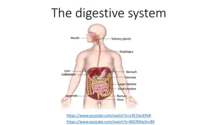
Parts of the Large Intestine Parts of the Large Intestine o (1,2,3,4,5) are found in the abdomen o (6,7) are found in the pelvis o (8) is found in the perineum Epiploic Appendices Characteristics of Colon Characteristics of Colon o NOT found in rectum & anal canal ONLY colon o Taeniae coli: Three longitudinal muscle bands o Sacculations (Haustra)= : Because the Taeniae coli are shorter than large intestine o Epiploic Appendices = : Short peritoneal folds filled with fat (Yellowish color) Anus or Anal canal
Peritoneal Covering Peritoneal Covering Parts devoid of (without) peritoneal covering Parts with mesentery1 Parts with Retroperitoneal2 1.Transverse colon3 2.Sigmoid colon 3.Appendix 4.Cecum 1.Ascending colon 2.Descending colon 3.Upper 2/3 of rectum 1.Lower 1/3 of rectum 2.Anal canal Rectum Anal canal 1- The peritoneum covers the anterior and posterior surfaces. 2- The peritoneum only covers the anterior surface 3- First 2 inches WITHOUT mesentery
Parts of the Large Intestine Parts of the Large Intestine Relation Relation *Transverse colon empty Stomach will be Superior *Transverse colon full Stomach will be Anterior Quadratus lumborum Anterior Posterior 1.Psoas major 2.Iliacus Cecum (Right) 1.Right kidney 2.Iliacus 3.Quadratus lumborum Ascending colon (Right) 1.Greater omentum 2.Anterior abdominal wall 3.Coils of small intestine (jejunum & ilum) 1.Psoas major 2.Iliacus 3.Quadratus lumborum 4.Left kidney Descending colons (Left) 1.Greater omentum 2.Anterior abdominal wall 3.Stomach* 1.Superior mesenteric vessels 2.2nd part of duodenum 3.Pancreas (head) Transverse colon Superior Inferior 1.Gallbladder 2.Stomach 3.Liver 1.Coils of small intestine (jejunum & ilum)
Colic flexures & Appendix Colic flexures & Appendix Colic flexures Splenic flexure (Left) Hepatic flexure (Right) Between Transverse colon & Spleen Position: higher | Angle: more acute Between Ascending colon & Liver Wider angle Appendix (Anatomically related to large intestine BUT Functionally related to immunity Lymphatic system ) The base of appendix is marked by McBurney s point*: A point at the junction of lateral 1/3 & medial 2/3 of a line traced from right anterior superior iliac spine to umbilicus. Surface Anatomy Extra Opening At posteromedial aspect of cecum, 1 inch below ileocecal junction 1.Retrocecal (most common) 2.Pelvic 3.Subcecal** 4.Preiliea 5.Postileal (least common) **Its original position but with development of the cecum, the position could change depend on equality of the developing sites of cecum, usually inequality causing shifting of appendix position (REMEMBER! The position of base not change) Positions *In case of appendicitis this area will have rebound tenderness (It refers to pain upon removal of pressure rather than application of pressure to the abdomen)
- Upper 1/3 of rectum is covered by peritoneum in front & side - Middle 1/3 of rectum is covered by peritoneum in front ONLY - Lower 1/3 of rectum & anal canal has no peritoneum cover Rectum Rectum Beginning as a continuation of sigmoid colon at level of S3 Continues as anal canal, one inch below & in front of tip of coccyx Its end is dilated to form the rectal ampulla Termination Length 13 cm (5 inches) Relations of Rectum in Pelvis Male pelvic Male pelvic 1.Posterior surfaces of urinary bladder 2.Seminal vesicles 3.Prostate gland Anterior 1.posterior wall of vagina Posterior 1.Sacral plexus 2.Sacrum 3.Coccyx Male pelvic Female pelvic
Relation between embryological origin of GIT Relation between embryological origin of GIT & & Supply Supply from esophagus proximal duodenum at major duodenal papilla (Celiac trunk) from distal duodenum after opening from cecum, appendix, ascending colon & right 2/3 of transvers colon (Superior mesenteric artery) Foregut Arterial Supply Midgut from left 1/3 of transvers colon, Descending colon, Sigmoid colon & Upper 2/3 of anal canal (Inferior mesenteric artery)* Hindgut Venous Drainage The veins of the gut form the tributaries of the portal vein which enters the liver and drains into the portal circulation (Portal vein liver sinusoid IVC) The lymph vessels follow the arteries. Ultimately, all the lymph is collected at the Preaortic lymph nodes (Superior & Inferior mesenteric). Lymph Drainage Ectoderm *lower 1/3 of anal canal | Somatic (inferior rectal from sacral plexus) Nerve Supply Midgut* *Endoderm | (Autonomic): Sympathetic + Vagus *Endoderm | (Autonomic): Sympathetic + pelvic splanchnic nerves (S2&S3) *It continues as the superior rectal artery in the root of the sigmoid mesocolon (pelvic region). Hindgut*
Cecum, Ascending & Descending Colons Peritoneal Covering Venous, Lymph & Nerve Supply Transverse Colon Appendix Rectum Venous drainage: Portal circulation Lymph drainage: Preaortic lymph nodes Nerve supply: Origin: Midgut Nerve supply: (Autonomic): Sympathetic + Vagus Origin: Hindgut Nerve supply: (Autonomic): Sympathetic + pelvic splanchnic nerves Origin: ectoderm (lower 1/3 of anal canal) Nerve Supply: Somatic (inferior rectal) Parts with mesentery: 1. Transverse colon 2. Sigmoid colon 3. Appendix 4. Cecum Retroperitoneal parts: 1. Ascending colon 2. Descending colon 3. Upper 2/3 of rectum Parts devoid of peritoneal covering: 1. Lower 1/3 of rectum 2. Anal canal Anterior relations: -Greater omentum -Coils of small intestine -Anterior abdominal wall Posterior relations: Cecum: 1. Psoas major 2. Iliacus Ascending colon: 1. Iliacus 2. Quadratus lumborum 3. Right kidney Descending colon: 1. Left kidney 2. Quadratus lumborum 3. Iliacus Colic flexures: 1-Hepatic flexure 2-Splenic flexure: higher + more acute angle Relations: Anterior: -greater omentum -anterior abdominal wall Posterior: 2nd part of duodenm, pancreas & superior mesenteric vessels Superior: -liver -gall bladder -stomach Inferior: coils of small intestine Surface anatomy: the base of appendix is marked by Mc Burney spoint Opening: At posteromedial aspect of cecum Positions: 1.Retrocecal most common 2.Pelvic 3.Subcecal 4.Preilieal 5.Postileal least common Beginning: at level of S3 Termination: continues as anal canal, one inch below & in front of tip of coccyx Length: 13 cm (5 inches) Relations: Posterior: sacrum, sacral plexus & coccyx Anterior: MALE PELVIS: seminal vesicles, posterior surfaces of urinary bladder & prostate gland FEMALE PELVIS: posterior wall of vagina
MCQs 1. The taeniae coli found in which of the following structure? A- Transverse colon B- Small intestine C- Rectum D- Anal canal 6. All the lymph in the GIT is collected at the: A- Preaorticlymph nodes (Superior & Inferior mesenteric) B- Preaorticlymph nodes (anterior & Inferior mesenteric) C- Postaorticlymph node D- Non of them 2. Which of the following part is with mesentery? A- Lower 1/3 of rectum B- Appendix C- Ascending colon D- Upper 2/3 of rectum 7. The termination of the rectum is: A- As a continuation of sigmoid colon at level of S3 B- Continues as anal canal, one inch below & in front of tip of coccyx C- Sacral plexus & coccyx D- A & C 3. Which of the following structure is an anterior relation of cecum? A- Psoas major B- lliacus C- Quadratus lumborum D- Coils of small intestine 8. Which one of the following parts of large intestine is found in the pelvis? A- Transverse colon B- Anal canal C- Rectum D- Cecum 4. The superior mesenteric vessels relate to Transverse colon? A- Anteriorly B- Posteriorly C- Superiorly D- Inferiorly 5. Which one of the following is the nerve supply of the Hindgut (endoderm): A- Sympathetic + pelvic splanchnic nerves B- Somatic (inferior rectal) C- Sympathetic + Vagus D- Non of them 9. Its surface anatomy is marked by Mc Burney s point: A- Rectum B- Colon C- Appendix D- Pancreas
Answers (1) A (2) B (3) D (4) B (5) A (6) A (7) B (8) C (9) C
SAQ 1. What are the characteristics of colon? 1) Taeniae coli 2) Sacculations 3) Epiploic appendices 2. What are the anterior relations of (Cecum Ascending & Descending colons): 1) Anterior abdominal wall 2) Coils of small intestine 3) Greater omentum 3. What are the posterior relations (Cecum Ascending & Descending colons): Cecum: 1. Psoas major 2. Iliacus Ascending colon: 1. Iliacus 2. Quadratus lumborum 3. Right kidney Descending colon: 1. Left kidney 2. Quadratus lumborum 3. Iliacus 4. Psoas major
Good luck Special thank for team436 Team Leaders: Faisal Fahad Alsaif Rawan Mohammad Alharbi Team Members: Abdulaziz Aldukhayel Abdulrahman Alduhayyim Rinad Alghoraiby Majd Albarrak References: 1.Girls & Boys Slides 2.Earthslab.com 3.TeachMeAnatomy.com Twitter.com/Anatomy437 Anatomyteam.437@gmail.com