Understanding the Anatomy and Histology of the Conjunctiva
This educational content explores the intricate details of the conjunctiva, a thin mucous membrane that lines the inner surface of the eyelids and globe. Topics include anatomical divisions, histological features such as epithelium and stroma, and common microbial conjunctival pathologies like bacterial, viral, and chlamydial conjunctivitis. Learning objectives encompass identifying clinical applications of conjunctival anatomy and histology, recognizing physical signs in conjunctival pathology, and understanding the management of various types of microbial conjunctivitis.

Download Presentation

Please find below an Image/Link to download the presentation.
The content on the website is provided AS IS for your information and personal use only. It may not be sold, licensed, or shared on other websites without obtaining consent from the author. Download presentation by click this link. If you encounter any issues during the download, it is possible that the publisher has removed the file from their server.
E N D
Presentation Transcript
The Conjunctiva The Conjunctiva Part Part 1 1 Dr. Zainab N. Hamoodi Lecturer ophthalmologist M.B.Ch.B. , F.I.C.M.S.(Ophth), ICO
Learning Objectives Learning Objectives To identify the clinical application of conjunctival anatomy & histology. To recognize the physical signs seen in conjunctival pathology. To recognize common types of microbial conjunctivitis (bacterial/viral/chlamydial) in terms of clinical course & management.
Contents : Contents : Anatomy Anatomy Conjunctival Conjunctival inflammation inflammation Microbial conjunctivitis : Microbial conjunctivitis : Bacterial conjunctivitis Bacterial conjunctivitis Viral conjunctivitis Viral conjunctivitis Chlamydial conjunctivitis Chlamydial conjunctivitis
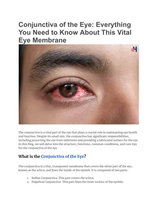
Anatomy : Anatomy : The conjunctiva is a thin transparent mucous membrane transparent mucous membrane lining the inner surface of the eyelids & surface of the globe as far as the Limbus Limbus . It can be divided into It can be divided into 3 1. 1. The Palpebral / Tarsal conjunctiva = lines the inner surface of the lids & firmly attached to it . 3 parts : parts : The Palpebral / Tarsal The Forniceal Forniceal conjunctiva 2. 2. The loose & redundant, may be thrown into folds , located at the fornix . conjunctiva = 3. The Bulbar Bulbar conjunctiva = covers the anterior sclera up to the limbus (covering the globe) .
Anatomical divisions Anatomical divisions
Anatomical divisions Anatomical divisions
Histology : Histology : 1. 1.Epithelium = Epithelium = Non keratinized stratified columnar/cuboidal epithelium about 2 - 5 cell layer in thickness. It contains goblet cells goblet cells (unicellular mucin secreting glands ) mucin is a component of the tear film .
2 2. . Stroma Stroma ( (substancia substancia propria propria) ) consists of richly-vascularized loose connective tissue, can be subdivided into: Superficial adenoid layer contains lymphoid tissue, it develops at 3 months after birth, hence inability of the neonates to develop follicular conjunctival reaction. Deep fibrous layer it contains accessory lacrimal glands. Inflammatory diseases damage the conjunctiva destruction of goblet cells/accessory lacrimal glands dry eye.
Conjunctivitis Conjunctivitis It is inflammation of the conjunctiva , which is either : Microbial OR Allergic Microbial OR Allergic Symptoms : Symptoms : Foreign body sensation, itching or burning sensation . Redness Discharge
Signs : Signs : Hyperemia = Hyperemia = dilatation of conjunctival blood vessels, non specific seen in microbial/allergic inflammation . Chemosis Chemosis = due to transudation of fluid from damaged blood vessels into subconjunctiva , non specific sign . = swelling of bulbar & fornix conjunctiva , Sub Sub- -conjunctival conjunctival hemorrhage = ocular trauma, viral conjunctivitis, or spontaneously . hemorrhage =
Conjunctival Conjunctival hyperemia hyperemia
Conjunctival Conjunctival chemosis chemosis
Papillae Papillae Multiple translucent hyperemic up growth of inflamed conjunctiva, non specific sign non specific sign = hyperplasia of epithelium + edema of stroma with central core of blood vessel .
Papillae Papillae ( (conjunctival conjunctival papillary reaction) papillary reaction)
Follicles Follicles Multiple solid nodular up growth pushing blood vessel aside = aggregation of inflammatory cells, lymphocytes & monocytes More specific More specific Viral infections Chlamydial infections drug hypersensitivity Infants less than 3 months old NO follicles NO follicles
Conjunctival Conjunctival follicles follicles
Follicles Follicles ( (conjunctival conjunctival follicular reaction) follicular reaction)
Membrane Membrane = coagulated exudates adherent to inflamed conjunctiva, seen in conjunctivitis + profuse secretion . True membrane True membrane = firmly adherent to inflamed conjunctiva , as in Diphtheria Pseudo membrane Pseudo membrane = loosely adherent to inflamed conjunctiva, as in gonococcal conjunctivitis
Membranes Membranes
Type of discharge Type of discharge Watery Watery viral/allergic Purulent Purulent severe bacterial conjunctivitis Mucopurulent Mucopurulent mild bacterial conjunctivitis Mucinous Mucinous severe allergic conjunctivitis
Regional Lymphadenopathy Regional Lymphadenopathy Involving the preauricular + submandibular L.N. viral, Chlamydia, gonococcal conjunctivitis Lab Ix : Lab Ix : Smears Gram stain, Giemsa stain Indicated Severe, chronic, follicular Ophthalmia neonatorum conjunctivitis
Microbial conjunctivitis Microbial conjunctivitis 1 1. Bacterial conjunctivitis . Bacterial conjunctivitis Common, acute self limiting infection, mainly caused by Strept. pneumoniae , Staph. aureus , Staph. epidermidis . Symptoms Symptoms Acute discomfort, irritation, F.B sensation . Frequently the eyelids are stuck together upon waking up in the morning, unilateral then becomes bilateral . Acute onset of redness, grittiness, discharge, Signs Signs Crusted eyelids Mucopurulent discharge Conjunctival congestion in the form of papillary reaction .
Treatment Treatment Local Local antibiotic eye drops &/or ointment Ciprofloxacin, Fucithalmic, Gentamicin ointment
Gonococcal Gonococcal conjunctivitis conjunctivitis Is caused by G-ve diplococci Neisseria gonorrhoea newborn (Ophthalmia neonatorum), young adults Symptoms Symptoms : : hyperacute hyperacute , profuse purulent profuse purulent ocular discharge . Signs Signs eyelid edema, erythema , conjunctival Chemosis In severe cases pseudo pseudo- -membrane Tender pre-auricular lymphadenopathy . Keratitis = preipheral corneal ulceration, may rapidly progress to perforation perforation within 48 hr = EMERGENCY Chemosis membrane
Management : Management : Hospital admission Hospital admission Topical antibiotics Topical antibiotics initially topical antibiotics should be given frequently every 5 minutes gradually reducing the frequency of instillation Systemic antibiotics Systemic antibiotics third generation cephalosprin (Cefotaxime)
Viral Conjunctivitis Viral Conjunctivitis Adenovirus Adenovirus is the most common Symptoms : Symptoms : F.B. sensation, redness, lacrimation Signs : Signs : Hyperemic conjunctiva, chemosis, watery sub conjunctival hemorrhage, follicular regional lymphadenopathy. watery secretion, follicular up-growth, Sometimes may affect the cornea keratitis keratitis = multiple intraepithelial gray granular dots cornea punctate epithelial punctate epithelial
Adenoviral Adenoviral keratoconjunctivitis keratoconjunctivitis Clinically 2 ocular syndromes caused by adenovirus : Pharyngoconjunctival Pharyngoconjunctival fever fever sero type 3 & 7 children with URTI Epidemic Epidemic keratoconjunctivitis keratoconjunctivitis sero type 8 & 9 keratitis > common , NO systemic symptoms
Adenoviral Adenoviral keratoconjunctivitis keratoconjunctivitis
Management Management Self limiting Self limiting disease, contagious contagious infection Transmission Transmission respiratory droplets/ ocular secretions/ contaminated towels or objects . Precautions Precautions washing hands + disinfecting ophthalmic instruments .
COVID-19 conjunctivitis Causative agent is SARS-CoV-2 (Severs Acute Respiratory Syndrome Corona Virus 2), well-known as COVID-19. Viral RNA recovered in tears of infected patients (including asymptomatic ones) so ocular surfaces serve as portal of entry & source for viral transmission. Conjunctivitis could be the first feature of the disease. Mechanism of infection exposure to aerosolized droplets or contaminated hand- eye contact.
Symptoms : Unilateral or bilateral red eye, lid swelling, watery eyes. Signs: Eyelid edema, conjunctival hyperemia, chemosis, watery discharge, follicular reaction, pre-auricular lymphadenopathy.
Diagnosis: Conjunctival swab analyzed for PCR negative results will not exclude the infection. Treatment: No therapeutic strategy against COVID-19 have been proved to be safe & effective to date. Precautions should be taken during examination of patients with features suggestive of viral conjunctivitis. Mandatory to inquire about respiratory symptoms/systemic review suggestive of potential COVID-19 infection.
Treatment: Inquire about contact with COVID-19 patients. Educating patients for meticulous eye-hand hygiene to avoid viral transmission. Symptomatic treatment for viral conjunctivitis & topical antibiotics for secondary bacterial conjunctivitis.
Chlamydia infection of the eye Chlamydia infection of the eye Trachoma Trachoma Adult inclusion conjunctivitis Adult inclusion conjunctivitis Ophthalmia Ophthalmia Neonatorum Neonatorum
Trachoma Trachoma Causative agent Chlamydia trachomatis serotype A,B,C serotype A,B,C Vector common fly common fly Leading cause of preventable blindness Leading cause of preventable blindness affects 500 million people Common in communities with poor hygiene Endemic Endemic in the middle east middle east region poor hygiene
Clinical Features Clinical Features Presentation : Presentation : In Bilateral redness, F.B. sensation, mucupurulent discharge Follicles : Follicles : mostly at upper palpebral conjunctiva upper palpebral conjunctiva + upper In 1 1st stdecade of life decade of life upper limbus limbus Intense inflammatory reaction Intense inflammatory reaction Kratitis Kratitis = Initially Initially punctate epithelial erosions (PEE) in upper cornea Pannus Pannus = sub epithelial fibro vascular ingrowth invading the cornea from above center = inflammation of the cornea
Scarring Scarring Follicles in upper palpebral conjuctiva disappear with scar as a stellate/horizontal line = Arlet Arlet s s lines lines Follicles in the upper limbus replaced by concave depressions = Herbert Herbert s pits s pits Complications Complications Lid scarring Lid scarring Trichiasis Trichiasis = posterior misdirection of eyelashes Entropion Entropion = inward inversion of the lid margin Conjunctival Conjunctival scarring glands/goblet cells Dry eye Cicatricial Cicatricial trachoma trachoma predisposes to microbial keratitis scarring destruction of accessory lacrimal Dry eye
TRACHOMA Follicles TRACHOMA Follicles
Corneal Corneal pannus pannus Trichiasis Trichiasis Entropion Entropion
Stages of trachoma = WHO classification Stages of trachoma = WHO classification TF = TF = Follicles in the upper palpebral conjunctiva TI = TI = intense inflammatory reaction TS = TS = Scarring TT = TT = Trichiasis TCO = TCO = Corneal opacification
Management : Management : SAFE strategy (WHO) SAFE strategy (WHO) S = S = Surgery for trichiasis A = A = Antibiotics for active disease F = F = Facial hygiene E = E = Environmental improvement Topical antibiotics Tetracycline eye ointment, twice daily for 6 weeks Systemic antibiotics Tetracycline/Doxycycline Young children systemic erythromycin OR Azithromycin 20 mg/kg single oral dose
Adult inclusion conjunctivitis Adult inclusion conjunctivitis Causative agent Chlamydia trachomatis serotype D to K STD typically affects young adults Presented Acute bilateral mucopurulent follicular conjuctivitis + regional LAP Keratitis; superior pannus may also occur. Treatment : Treatment : Topical Topical antibiotics Tetracyclin eye ointment twice daily for 6 weeks Systemic Systemic antibiotics tetracyclin/doxycycline Pregnant women systemic erythromycine OR Azithromycin 20 mg/kg single oral dose
Adult chlamydia inclusion conjunctivitis Adult chlamydia inclusion conjunctivitis
References : 1. Kanski Clinical Ophthalmology 2. Oxford handbook of clinical ophthalmology 3. American Academy of Ophthalmology