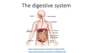
Histology Overview of Digestive and Urinary Tracts in Pharmacy Practice

This histology practice session delves into the intricate details of the digestive and urinary tracts studied in the 8th week of Pharmacy school. Explore images showcasing the structures of the colon, vermiform appendix, liver, pancreas, and nephron. Gain insights into the histological features of these organs, including epithelial cells, glandular tissues, sinusoids, lobules, and more.

Download Presentation

Please find below an Image/Link to download the presentation.
The content on the website is provided AS IS for your information and personal use only. It may not be sold, licensed, or shared on other websites without obtaining consent from the author. Download presentation by click this link. If you encounter any issues during the download, it is possible that the publisher has removed the file from their server.
E N D
Presentation Transcript
Digestive tract II. Urinary tract Faculty of Pharmacy 8th week histology practice Department of Anatomy, Histology and Embriology
Colon Simple coumnar epith. with many goblet cells Lieberk hn cripts in the l. propria Muscularis mucosae Inner circular&outer longitudinal layers Submucosa NO glands Muscularis externa Inner circular&outer longitudinal layers the outer layer is not continous, it forms the 3 tenia coli Serosa OR Adventitia
Vermiform appendix Lymph nodules in the lamina propria and submucosa Tenia coli is absent
Liver Hepatic lobules hexagonal shape (portal lobule, acinus) Portal triad portal vein, artery, bile duct Hepatic sinusoids between the hepatic chords Central vein
Hepatic lobule Central vein Hep. sinus Hep. chord Interlobular vein Portal Interlobular artery triad Interlobular duct
Central vein Portal triad
Interlobular duct Interlobular vein Interlobular artery Portal triad
Histology of the pancreas Serous gland Centroaciner cell Langerhans islets endocrine part
Macula densa Malphigian corpuscule interdigitating podocytes
Macula densa Proximal tubule Parietal layer of Bowman s capsule Distal tubule
72. Liver (HE) Central vein Portal triad
72. Liver (HE) Interlobular duct Interlobular vein Interlobular artery Portal triad
74. Kidney (HE) Macula densa Proximal tubule Parietal layer of Bowman s capsule Distal tubule


")
")
")