Renal Block Pathology Practical: Anatomy and Histology Overview

The Renal Block Pathology Practical provides a comprehensive insight into the anatomy, structure, and histology of the kidney nephrons, including normal and abnormal conditions such as acute kidney injury. The session covers topics like the structure of the nephron, normal kidney gross anatomy, renal corpuscle histology, cortical tubules, and the causes of acute kidney injury. Detailed images and descriptions enhance the understanding of renal pathology.

Download Presentation

Please find below an Image/Link to download the presentation.
The content on the website is provided AS IS for your information and personal use only. It may not be sold, licensed, or shared on other websites without obtaining consent from the author. Download presentation by click this link. If you encounter any issues during the download, it is possible that the publisher has removed the file from their server.
E N D
Presentation Transcript
RENAL BLOCK PATHOLOGY PRACTICAL I & II Prof. Ammar Al Rikabi Dr. Sayed Al Esawy Dr. Marie Mukhashin Dr. Shaesta Zaidi Prepared by: Head of Pathology Department: Dr. Abdulmalik Al Sheikh
Anatomy of the Kidney Pathology Dept , KSU Renal Block
NEPHRON STRUCTURE Pathology Dept , KSU Renal Block
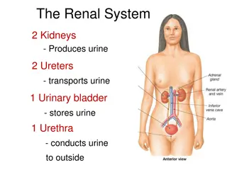
KIDNEY ANATOMY : NEPHRONS Nephron is the functional unit of the kidney. Each kidney contains about 1,000,000 to 1,300,000 nephrons. The nephron is composed of glomerulus and renal tubules . The nephron performs its function by ultra filtration at glomerulus and secretion and reabsorption at renal tubules. Pathology Dept , KSU Renal Block
Normal Kidney - Gross In cross section, this normal adult kidney demonstrates the lighter outer cortex and the darker medulla, with the renal pyramids into which the collecting ducts coalesce and drain into the calyces and central pelvis. Pathology Dept , KSU Renal Block
Renal Corpuscle Normal Histology Pathology Dept , KSU Renal Block
Renal Corpuscle Normal Histology Normal glomerulus by light microscopy. The glomerular capillary loops are thin and delicate. Endothelial and mesangial cells are normal in number. The surrounding tubules are normal Pathology Dept , KSU Renal Block
Normal Cortical Tubules Normal cortical tubules, interstitium, and peritubular capillaries; most of the tubules are proximal, with well-defined brush borders (PAS stain). Pathology Dept , KSU Renal Block
PRACTICAL SESSION : 1 ACUTE KIDNEY INJURY Pathology Dept , KSU Renal Block
Acute Kidney Injury Causes: Pre-renal (All those that decrease effective blood flow to the kidney) Low blood volume, low blood pressure, and heart failure. Renal artery stenosis, and renal vein thrombosis. Renal ischemia. Renal: Glomerulonephritis (GN). Acute tubular necrosis (ATN). Acute interstitial nephritis (AIN). Post-renal: (is a consequence of urinary tract obstruction) Benign prostatic hyperplasia. Kidney stones. Obstructed urinary catheter. Bladder stone . Bladder, ureteral or renal malignancy. Pathology Dept , KSU Renal Block
Acute Kidney Injury Kidney showing marked pallor of the cortex, contrasting to the darker areas of surviving medullary tissue. Pathology Dept , KSU Renal Block
Acute Tubular Necrosis Acute tubular necrosis is manifest by vacuolated cells and sloughed, necrotic cells in tubular lumina, with some tubules lined by flattened epithelium and some showing frank necrosis Pathology Dept , KSU Renal Block
Acute Interstitial Nephritis There is edema in addition to preexisting mild tubulointerstitial fibrosis . There is a prominent interstitial eosinophilic component, in addition to lymphocytes and plasma cells Pathology Dept , KSU Renal Block
POLYCYSTIC KIDNEY Pathology Dept , KSU Renal Block
Normal vs Polycystic Kidney Pathology Dept , KSU Renal Block
Polycystic kidney Gross Anatomy Markedly enlarged kidney and replacement of the renal parenchyma by numerous cysts of variable sizes in entire cortex Pathology Dept , KSU Renal Block
Gross Polycystic kidney and its Cut Section Cut surface of the kidney, showing extensive cortical destruction by cysts Massively enlarged kidney disrupted by numerous cysts Pathology Dept , KSU Renal Block
Autosomal dominant Polycystic Kidney Disease Variable sized cysts fill most of the parenchyma No glomeruli seen. COMPLICATIONS: Hypertension, Renal failure
Infantile Polycystic kidney Gross Coronal section of an infantile polycystic kidney Pathology Dept , KSU Renal Block
Polycystic kidney Histopathology Kidney of child with autosomal dominant PCKD. a. Coronal section of a polycystic kidney. b. histology demonstrating glomerular cysts . Note the normal-sized glomeruli with the enlarged Bowman s space and tubular cystic changes Pathology Dept , KSU Renal Block
PRACTICAL SESSION : 2 INFECTION OF THE URINARY TRACT Pathology Dept , KSU Renal Block
ACUTE (POST-STREPTOCOCCAL) GLOMERULONEPHRITIS Pathology Dept , KSU Renal Block
Acute (Post-streptococcal) Glomerulonephritis This glomerulus is hypercellular and capillary loops are poorly defined. This is a type of proliferative glomerulonephritis known as post- infectious glomerulonephritis Pathology Dept , KSU Renal Block
Acute (Post-streptococcal) Glomerulonephritis The hypercellularity of post-infectious glomerulonephritis is due to increased numbers of epithelial, endothelial, and mesangial cells as well as neutrophils in and around the glomerular capillary loops Pathology Dept , KSU Renal Block
Acute (Post-streptococcal) Glomerulonephritis High power LM of a hypercellular glomerulus; numerous capillaries contain inflammatory cells, mostly neutrophils Pathology Dept , KSU Renal Block
Acute (Post-streptococcal) Glomerulonephritis Section of the kidney shows: The glomeruli are enlarged, lobulated and hypercellular with obliteration of capsular space. Cellularity is due to proliferation of endothelial and mesangial cells with some neutrophils. Many capillaries appear obliterated. Tubules show degenerative changes. Pathology Dept , KSU Renal Block
ACUTE & CHRONIC PYELONEPHRITIS Pathology Dept , KSU Renal Block
Pyelonephritis with small cortical abscesses Pyelonephritis with small cortical abscesses Pathology Dept , KSU Renal Block
Classic picture of Pyelonephritis This kidney is bisected to reveal a dilated pelvis and calyxes filled with a yellow- green purulent pus which is consistent with a pyelonephritis. The cortex and medulla are pale and the corticomedullary junction is ill-defined. No tumors are seen. Pathology Dept , KSU Renal Block
Acute Pyelonephritis - Histopathology Acute pyelonephritis is diagnosed by intratubular aggregations of polymorphonuclear neutrophils (PMNs). There may be surrounding interstitial inflammation with a mixture of PMNs, lymphocytes, and plasma cells, but the predominant inflammation is within the tubule Pathology Dept , KSU Renal Block
Acute Pyelonephritis - Histopathology Numerous PMN's are seen filling renal tubules across the center and right of this picture. These leukocytes may form into a cast within the tubule. Casts appearing in the urine originate in the distal renal tubules and collecting ducts Pathology Dept , KSU Renal Block
Chronic Pyelonephritis - Gross Pathology The picture shows slightly atrophic and deformed kidneys with cortical coarse scars . Pathology Dept , KSU Renal Block
Chronic Pyelonephritis - Histopathology This is chronic pyelonephritis where a large collection of chronic inflammatory cells . The severity of disease depends upon the amount of remaining functional renal parenchyma Pathology Dept , KSU Renal Block
Chronic Pyelonephritis - Histopathology High power shows periglomerular fibrosis , glomerular sclerosis and hyalinization with marked chronic interstitial inflammation . Pathology Dept , KSU Renal Block
Chronic Pyelonephritis - Histopathology -Glomeruli show varying degrees of sclerosis & periglomerular fibrosis. -Tubules show varying degrees of atrophy, Some tubules are dilated and filled with Eosinophilic hyaline casts resembling colloid (thyroidization). -Interstitial tissue shows chronic inflammatory cells infiltrate and fibrosis. Pathology Dept , KSU Renal Block
HYDRONEPHROSIS Pathology Dept , KSU Renal Block
Hydronephrosis The most common causes are: Foreign bodies like calculi with obstruction, Atresia of the urethra, Benign prostatic hyperplasia , Neoplasia of the prostate and bladder Spinal cord damage with paralysis of the bladder . The picture shows markedly dilated renal pelvis and calyces with atrophic and thin renal cortex /parenchyma Pathology Dept , KSU Renal Block
Hydronephrosis Complications of hydronephrosis --Urinary tract infection --Pyelonephritis. --Renal failure. Bisected kidney shows markedly dilated renal pelvis and calyces with atrophic and thin renal cortex /parenchyma Pathology Dept , KSU Renal Block
Hydronephrosis Markedly dilated renal pelvis and calyces with atrophic and thin renal cortex Pathology Dept , KSU Renal Block
PRACTICAL SESSION : 2 NEPHROTIC SYNDROME Pathology Dept , KSU Renal Block
Membranous Glomerulonephritis Membranous glomerulonephritis ( The common cause of Nephrotic syndrome in adults): the capillary loops are thickened and prominent, but the cellularity is not increased. Pathology Dept , KSU Renal Block
Membranous Glomerulonephritis Close-up of glomerulus illustrating rigid, uniformly-thickened capillary walls Pathology Dept , KSU Renal Block
Membranous Glomerulonephritis The thickened capillary wall shows numerous "holes" in tangential sections, indicating deposits. (Deposits do not take up the silver stain.) Well-developed spikes around the deposits are not present here. Pathology Dept , KSU Renal Block
NEPHRITIC SYNDROME (RPGN) Pathology Dept , KSU Renal Block
Rapid Progressive Glomerulonephritis (RPGN) Gross appearance of RPGN - note the flea beaten appearance Pathology Dept , KSU Renal Block
Rapid Progressive Glomerulonephritis (RPGN) Seen here within the glomeruli are crescents composed of proliferating epithelial cells. Crescentic glomerulonephritis is known as rapidly progressive glomerulonephritis (RPGN) because this disease is very progressive Pathology Dept , KSU Renal Block
Rapid Progressive Glomerulonephritis (RPGN) Epithelial cells of Bowman capsule are proliferated . Infiltrating WBCs such as monocytes and macrophages also proliferate compressing the glomerulus, forming a crescent-shaped scar Pathology Dept , KSU Renal Block


")
 Glomerulonephritis")
 Glomerulonephritis")
 Glomerulonephritis")
 Glomerulonephritis")


")
")
")