Introduction to Histology and Cell Structure

Histology is the microscopic study of normal tissues utilizing light and electron microscopes. This field explores the composition and function of cells, focusing on the nucleus, cytoplasm, organelles, and inclusions. Thin tissue sections stained with Haematoxylin and Eosin reveal distinct cellular components like the deeply basophilic nucleus and chromatin. The nucleus, comprising the nuclear envelope, chromatin, nucleolus, and nucleoplasm, plays a vital role in cellular vitality, genetic information storage, and RNA synthesis. Understanding these cellular structures is essential in histological analysis.

Download Presentation

Please find below an Image/Link to download the presentation.
The content on the website is provided AS IS for your information and personal use only. It may not be sold, licensed, or shared on other websites without obtaining consent from the author. Download presentation by click this link. If you encounter any issues during the download, it is possible that the publisher has removed the file from their server.
E N D
Presentation Transcript
Lecture 1 : Introduction to Histology and Cell Structure - Colour index : Red : important Grey : doctors notes
Objectives : What is histology and how it is studied? Composition of the cell: Light microscopic (L/ M) and electron microscopic (E/M) . Function of each component: - Nucleus. - Cytoplasm : - Organelles: membranous and non membranous. - Inclusions.
Introduction : Histology : is the microscopic study of normal tissues. Types of microscopes: LM & EM Cells Tissues Organs Systems Thin sections are cut and mounted on glass slides. Sections are stained with Haematoxylin (H) and Eosin (E). *Nucleus is always (basophilic) blue *Cytoplasm could be acidophilic (red) or basophilic (blue) LM : Produce colourful images . EM : Produce black and white images .
Nucleus (L/M) Shape of nuclei : The Cell It Is the structural & functional unit of all living tissues. Cells have different shapes & size. The Cell is made of: 1- Nucleus 2- Cytoplasm
Appearance of nuclei : Dark Nucleus (Deeply-stained Nucleus) Deeply Basophilic Nucleus Vesicular (Open Face) Nucleus.
The Nucleus Formed of: 1. 2. 3. 4. Nuclear envelope Chromatin Nucleolus Nucleoplasm Function of the Nucleus : It is essential for the vitality and division of the cell. It is the site of storage of genetic information. It is the site of formation of the three types of RNA.
1- Nuclear envelope Definition: A double membrane with many pores surroundings the Nucleus . Formed of : 1- Outer membrane 2- Inner membrane 3- Nuclear pores ( provide communication between nucleus and cytoplasm) * the only double membrane surrounded structures in the cell are Nucleus and Mitochondria . 2- Chromatin -Functions of chromatin : Carries genetic information . Directs protein synthesis . -Formed of DNA and protein Two forms : Euchromatin : extended active chromatin electron-lucent areas =(pale) Heterochromatin : condensed inactive chromatin electron-dense areas (dark)
3- Nucleolus E/M : appear mostly as dark mass (electron-dense) (the largest one). L/M : appear as spherical dark basophilic mass. Usually one . Not surrounded by a membrane . Function: formation of ribosomal RNA (rRNA) witch responsible for protein synthesis in the cytoplasm . 4- Nucleoplasm It is a clear fluid medium in which all the contents of the nucleus are embedded. Function: Provides a medium for movement of 3 types of RNA (ribosomal , messenger and transfer RNA) from the nucleus to the cytoplasm.
Sex chromatin (Barr body) : A dark stained mass of chromatin , usually adherent to the inner aspect of the nuclear envelope of female somatic cells e.g. Buccal epithelial cells. A drumstick mass protruding from the nucleus of neutrophils. Represents one of the two X chromosomes which is inactive (condensed) in normal female (heterochromatin). Seen in normal female cells. Absent in normal males cells . Absent in females with Turner s syndrome XO. Seen in males with Klinefelter s syndrome XXY . Extra Barr body (drumstick)
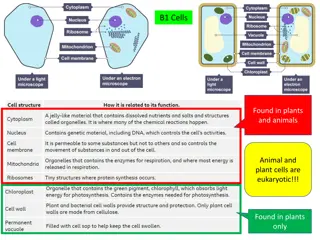
o Cytoplasm : is formed of: Organelles: They are specialized structures , essential for vital processes of the cell. Inclusions: They are not essential for vitality of cells , may be present or absent. Examples are lipids , glycogen and pigments like melanin & lipofuscin. A) Membranous : B) Non-Membranous : o Cytoplasmic organelles : cell membrane. mitochondria . endoplasmic reticulum. Golgi apparatus. lysosomes . secretory vesicles . Ribosomes. Centrioles . cilia and Flagella . Filaments (actin, myosin, intermediate filaments). Cytoskeleton (Actin, Intermediate filaments, Microtubules).
A) Membranous : How does it appear ? Cell membrane: A very thin membrane surrounding the cell. Function : Selective barrier. Chemical structure: - Phospholipid molecules : arranged in two layers. - Protein molecules: peripheral and integral proteins. - Carbohydrate molecules: attached to either proteins or lipids (glycoproteins and glycolipids) forming the surface or cell coat. LM (Light Microscope) EM (Electron Microscope) 2 dark lines separated by a light line (Trilaminar appearance). Not visible. Function of (Glycocalyx). 1- protection of the cell 2- cell recognition and adhesion.
Specializations of Cell Membrane : Cilia: Long motile hair-like structures surrounded by cell membrane. Their core is made of microtubules. Microvilli (Brush border) : Cylindrical cytoplasmic projection of apical surface to increase surface area . Their core contains actin filaments. Intercellular junctions 1. Occluding (Tight) Junction: seals the intercellular space. 2. Adherening Junction: fixes adjacent cells together: a) Zonula Adhering Junction. b) Desmosome (Macula Adherening Junction). 3. Gap junction: Allow free communication between the cells. *When a combination of 1 , 2a and 2b is present , this is called a junctional complex. 1 2a 2b 3
Mitochondria: - Each mitochondrion is rod-shaped . - The wall is composed of 2 MEMBRANES. - The outer is smooth, the inner is folded to form CRISTAE. - The cavity is filled with mitochondrial matrix, which contains enzymes. Also contains it s own DNA . Function: 1) Generation of ATP , which is the source of energy for the cell , They are called the power-house of the cell. 2) They can form their own proteins and undergo self replication.
Endoplasmic reticulum It is a system of communicating membranous tubules, vesicles, and flattened vesicles (cisternae). Rough Endoplasmic Reticulum Smooth Endoplasmic Reticulum Membranous sheets of flattened tubules & vesicles WITH ribosomes on the surface. Membranous tubules and vesicles , with NO ribosomes on the surface. Function: 1) Synthesis of proteins by ribosomes on its outer surface. 2) Transfer vesicles , transfer the formed protein to Golgi. Function: 1) Synthesis of lipids & cholesterol. 2) Synthesis of steroid hormones, e.g. cortisone. 3) Helps muscle contraction, by acting as a calcium pump. 4) Detoxification of drugs & toxins. FROM 437 TEAMWORK
Secretory vesicles Golgi apparatus The secretory apparatus of the cell. Consists of stacked saucer-shaped flattened vesicles. Each vesicle has two faces: 1- Convex (FORMING) face: receives transfer vesicles. 2- Concave (MATURE) face: forms secretory vesicles. Mature face Transfer vesicles Forming face Functions: 1- Sorting, modification & packaging of proteins. 2- Secretory vesicles formation. 3- Formation of lysosomes.
Lysosomes The digestive apparatus of the cell. E/M: Spherical membranous vesicles. Contain hydrolytic enzymes. Originate from mature surface of the Golgi apparatus, while their hydrolytic enzymes are formed in the rough endoplasmic reticulum. Function: intracellular digestion of ingested material or old organelles.
B) Non-Membranous Ribosomes LM: Basophilic cytoplasm is due to numerous ribosomes. Consist of ribosomal RNA (rRNA), combined with proteins. EM: Formed of 2 subunits. Free in the cytoplasm (may form polyribosomes) or attached to rER Formed in the nucleolus. Function: Protein synthesis.
Microtubules-containing organelles : Centrioles Cilia Flagella - 2 cylinders, perpendicular to each other . - Wall is made of 9 triplets of microtubules, i.e. 27 microtubules. - Hair-like striations on the free surface of some cells. - Basal body is similar to centriole. - Shaft is formed of 9 doublets and 2 central singlets of microtubules, i.e. 20 microtubules. Function: Movement of particles or fluids on the free surface of the cell in one direction. - Longer and larger than cilia. - Form the tails of sperms. Functions: 1- Essential for cell division. 2- Formation of CILIA and FLAGELLA. Function: Important for movement of the sperms. FROM 437 TEAMWORK
Microfilaments Cytoskeleton : It is the structural skeleton of the cell. Consists of: 1- Microfilaments (actin). 2- Intermediate filaments, e.g. Keratin. 3- Microtubules. Functions: 1- Maintains shape of the cell. 2- Helps transport of material within the cell. Microtubules Intermediate filaments
o Clinical application: Immotile cilia syndrome: Disorder that causes infertility in male and chronic respiratory tract infection in both sexes. caused by immobility of cilia and flagella induced by deficiency of dynein. Dynein protein is responsible for movements of cilia and flagella.
MCQs : 1- Which organelle sorts and packages proteins within a cell? A) Rough endoplasmic reticulum B) Smooth endoplasmic reticulum C ) Lysosome D) Golgi apparatus 5- In Golgi apparatus Which face receiving transfer vesicles ? A) Convex face B) Concave face C) Both D) Neither 2- Which organelle is considered the powerhouse of the cell? A) Rough endoplasmic reticulum B) Smooth endoplasmic reticulum C) Mitochondrial D) Lysosome 6- Smooth endoplasmic reticulum is the site of A) protein synthesis B) carbohydrate synthesis C) amino acid synthesis D) Lipid synthesis 3- Which of the following is NOT a membranous organelle? A )microtubules B )lysosomes C )mitochondria D ) endoplasmic reticulum 7- If you see the cytoplasm in microscope with blue color (basophilic), that means the cytoplasm stained with? A ) Hematoxylin(H) b ) Aldehyde fuchsine C ) Cresyl violet D) Eosin(E) 4- Microfilaments are composed mainly of a protein called A ) tubulin B ) actin C ) myosin D ) Chitin 8- If you see electron-lucent areas (pale) in microscope, that means the form of chromatin is ? A ) Euchromatin B) Heterochromatin C) A & B D ) Barr body
Team members : Alhanouf alhaluli Rawan alzayed Renad alkanaan Nouf albrikan Roaa aljohani Abdullah alassaf Abdullah altuwaijri Talal jamal aldeen Faisal alqifari Team leaders : Noura alnasser Abdullah shadid


")


 :")


 Membranous :")


 Non-Membranous")