Understanding Tissues: Types, Characteristics, and Functions

Explore the intricate world of histology and tissues, where groups of cells with similar structures and functions come together to support various bodily processes. From epithelial to nerve tissues, delve into the differences in tissue classes, cell types, and extracellular matrices. Discover the unique features of cardiac muscle, bone tissue, and skeletal muscle patterns, unraveling the essential role each plays in maintaining the body's structural integrity and function.

Download Presentation

Please find below an Image/Link to download the presentation.
The content on the website is provided AS IS for your information and personal use only. It may not be sold, licensed, or shared on other websites without obtaining consent from the author. Download presentation by click this link. If you encounter any issues during the download, it is possible that the publisher has removed the file from their server.
E N D
Presentation Transcript
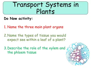
Tissues Groups of cells similar in structure and function - the structure plays a role into the function The structure and function complement each other
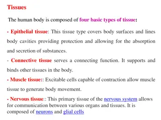
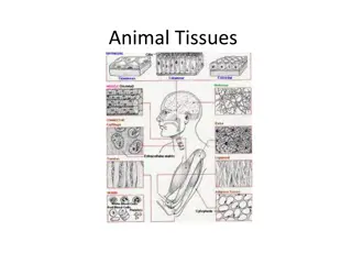
Types of Tissues Epithelial tissue Connective tissue Muscle tissue Nerve tissue Atoms to Molecules to Cells
Tissue Characteristics Differences between the tissue classes Types and functions of cells Characteristics of matrix (extracellular matrix) Fibrous proteins Ground substance thick or thin Clear gels (ECF, tissue fluid, interstitial fluid, tissue gel) Rubbery or stony in cartilage or bone
Tisssue Characteristics Space occupied by cells versus matrix Connective tissue cells are widely separated Little matrix between epithelial and muscle cells
Cardiac Muscle - one nucleus in cardiac muscle
Skeletal muscle - alternating dark and white patterns in skeletal muscles
Smooth Muscle - they do not have alternating dark and light muscles
Epithelial Tissue Two main types - Covering and lining epithelia - on external and internal surfaces Glandular epithelia Secretory tissue in glands
Glandular Epithelia ge glandular epithelia lp lamina propria
Epithelial Tissue Layers of closely adhering cells Flat sheet with upper surfaces exposed to the environment or an internal body cavity No blood vessels Underlying connective tissue supplies oxygen Rests on basement membrane Thin layer of collagen and adhesive proteins Anchors epithelium to connective tissue
Basement Membrane Not only an anchoring gel but also provides nutrients to the epithelium
Squamous covering Cuboidal and Columnar absorption The nucleus of columnar cells are found on the lower third of a cell
Simple Squamous 1 layer of flat cells
Serous Membranes watery membrane
Small intestine has epithelial tissue lining the inside and outside Outside of small intestine serous membrane Outer layer of the heart serous membrane What is the common feature of the small intestine and the heart?
Heart is constantly pumping the serous membrane also reduces wear and tear Small intestine produces watery secretions - the water in the serous membrane reduces friction and therefore reduces wear and tear
Surrounding the alveoli air sacs are capillaries Alveoli capillaries for oxygenation and removal of carbon dioxide Allows for rapid exchange if you have simple squamous epithelium
Simple Cuboidal Epithelium Found in areas of secretion and excretion Kidneys
Simple Cuboidal Epithelium One layer of cube-shaped cells
Simple Columnar Epithelium Tallest of the epithelial cells Found in small intestines almost all of absorption of nutrients is found here
Villi in the apical surface are microvilli
Microvilli they form the brush border Small extensions of the cell
The taller the cells the higher the chance of absorption The microvilli increases the absorptive area by increasing the height of cells Whenever you see cells with microvilli you should think that these cells are used to absorb everything
Pseudostratified Columnar Epithelium Stratified multi-layered - every single cell originates on one exact membrane but they are of different heights that is why they look stratified Respiratory mucosa
Ciliated Epithelium respiratory mucosa - lines the conducting airway of the lungs
Respiratory Mucosa Goblet Cells Secrete mucus and line the conducting airways Very sticky secretion Release a protein called Mucin Mucin combines with water and it becomes mucus We want to clear the air before it reaches the our air sacs so that it would not damage our lungs
Stratified Squamous Epithelium 2 major types a. keratinized b. non-keratinized
Stratified Squamous Non Keratinized wet areas of the body