Understanding Epithelial Tissues: Classification and Cellular Characteristics

Epithelial tissues are classified based on their structure and organization, with various types exhibiting distinct features like stacking or lack of stacking. These tissues are held together by special attributes, contributing to their functions within organs and systems. By examining tissues under a microscope, we can observe different cell types and their embryonic origins, shapes, activities, and surrounding matrices. Understanding these classifications aids in identifying tissue-related diseases and their implications. The angle of observation and histological processes impact how tissues are viewed and interpreted.

Download Presentation

Please find below an Image/Link to download the presentation.
The content on the website is provided AS IS for your information and personal use only. It may not be sold, licensed, or shared on other websites without obtaining consent from the author. Download presentation by click this link. If you encounter any issues during the download, it is possible that the publisher has removed the file from their server.
E N D
Presentation Transcript
How are epithelial tissues classified and held together? 9/16 This lecture content will be on Test#2, not Mondays test #1 Chapter 5: Chapter 5: 1) What is a tissue? 1) What is a tissue? 2) What is the embryonic pattern of development that creates different 2) What is the embryonic pattern of development that creates different tissues? tissues? 3) How do we view the things we see with a microscope? 3) How do we view the things we see with a microscope? 4) How are epithelial cells classified? 4) How are epithelial cells classified? 5) Four types lack stacking 5) Four types lack stacking 1 1- - Simple Simple squamous squamous ET characteristics ET characteristics 2 2- - Simple Simple cuboidal cuboidal ET characteristics ET characteristics 3 3- - Simple columnar ET characteristics Simple columnar ET characteristics 4 4- - Pseudostartified Pseudostartified ET characteristics ET characteristics 6) Four types have stacking 6) Four types have stacking Stacking gives a tissue special attributes Stacking gives a tissue special attributes Sign Up outside the AP lab for your special Lab Exam time for next Wed or Sign Up outside the AP lab for your special Lab Exam time for next Wed or Thur Thur .only one time per person. .only one time per person.
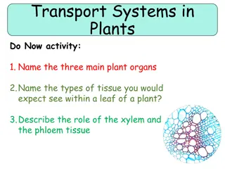
How do we classify/describe the different cell types that make up tissues, organs, organ systems and the body? Tissue : a set of cells with similar appearance and function together in an organ. How do we consider tissues? 1) Embryonic Origin: what cells were the embryonic precursors? 2) Shape/Appearance: are cells round, flat or cube etc? 3) Things leaving cells: Secretion vs. Excretion: S: material has physiological function E: waste removal from cell/body 4) Activity: Do cells use contraction, exocytosis, endocytosis or phagocytosis? 5) Matrix: What extracellular material is observed around the cells? Collagen, Elastin, Cartillage, Interstitial Fluids These factors let us classify the tissue type ! Tissues classification lets us understand the cause/effects of diseases.
How does the angle of observation affect what we observe? Two dimensional interpretation of three dimensional objects can be very difficult. Terms used to describe orientation of tissue section: Longitudinal View vs. Cross Sectional View Oblique View: Mix of the two for cells/tissue Histological fixing and sectioning of tissue Object on Slide Histological Staining of tissue Colors on Slide Sometimes fixation removes a part of the original tissue and what is seen on the slide is the remnant of where the cell used to be. The color we observe on a slide is usually created by the stains the tissue was prepared with. Try to interpret what you see on a slide as what it was .
What are our 3 Primary Germ Layers? Why are germ layers improtant? #1: Life begins as a fertilized egg (zygote; Week 1) that begins to divide. #2: Life progresses to a hollow ball of cells with a yolk sac underneath (blastoceol; Week 2) Outside surface= ecto derm Inside surface next to yolk sac= endo derm #3: Week2-3 The ball of cells forms a neural tube (CNS). Cells squeeze between the edges and migrate into the middle space between the yolk sac and the outside.
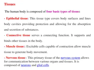
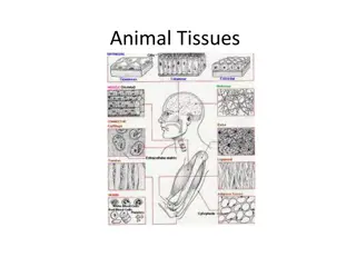
How do the three primary germ layers create the four primary classes of tissue? 1) Ectoderm: Epithelial Tissues Becomes: epidermis, hair, glands of skin, nerves, brain, a few blood vessels, and spinal cord Basement membrane holds them to underlying base! 2) Endoderm: Epithelial Tissues Becomes lining of lungs and glands of the gut 3) Mesoderm: Middle Tissues Becomes: dermis of skin, muscle, blood and connective tissues Remember that cells can migrate in the body during embryonic development. These 3 germ layers create the 4 Primary Tissue Classes: 1) Epithelial 2) Connective 3) Nervous 4) Muscular
The most fundamental way to organize epithelial cell classes is by layer and cellular shape (Know Table 5.2). Four types have a single epithelial cell layer: Simple Squamous Simple Cuboidal Simple Columnar Psuedostratified Columnar Four types have layered epithelial cells: Stratified Squamous 1)-with the protein keratin 2)-without the protein keratin 3) Stratified Cuboidal 4) Transitional Why is the basement membrane so important? This is cellular super-glue for all epithelial tissues! Why are underlying connective tissues required?
Why is the shape of simple squamous epithelium important for its function? Where is SSE: alveoli, blood vessels, serous membrane of organs, glomerular capsule Key Characteristic: FLAT and THIN! Typical packing and desmosomes : Why do we need a smooth surface? Why do we need a thin barrier/membrane? Where does gas exchange occur? Why does filtration occur? SSE tissues lining all blood vessels consist of endothelial cells Additional Endothelial Functions Additional Endothelial Functions: Hormonal functions- Contraction, inflammation and permeability- Clot Prevention-
Simple cuboidal epithelium helps with: protection, secretion and reabsorption. Key characteristic: CUBE SHAPED! Tight packing: Desmosomes: Provides Glandular Functions (Secretions): ovary, salivary, thyroid, pancreas, and liver Kidney has dual functions: Absorption and Secretion-
How do we protect cells and underlying tissues in places, such as the stomach? How do we prevent acids, abrasives, or pathogenic organisms from damaging underlying tissues? Simple Columnar Epithelium Simple Columnar Epithelium: Longer than they are wide to put nuclei by basement membrane for extra safety: Tough basement membrane: SCEs secrete digestive enzymes: SCEs help with nutrient absorption: Some have cilia (uterine tubes): Huge intestinal surface area with villi (cells) and microvilli (cell structures)= nutrient/water absorption Specialized Goblet cells make mucus to lubricate the passage of materials
Pseudostratified Columnar Epithelium (PCE) has cilia that move materials embedded in mucus in a single direction. Why is it called pseudostratified? Nuclei are at several different levels in cell- All cells still reach basement membrane- It only appears multi-layered Goblet cells are important! Importance of cilia for the airways? Importance of cilia in male reproductive tract? Importance of basement membrane?
Stratified Epithelial cells (Table 5.3) can be: 1) Squamous with keratin 2) Squamous without keratin, 3) Cuboidal or 4) Transitional. Callus on Sole of Foot: SSE-K...creates a nasty water insoluble place where bacteria can t live and water can t evaporate from the body! why? Mucosa of vagina, mouth, anus, esophogus are covered by: SSE-NoK why? Ovarian follicles and seminiferous tubules of the testes: SCE why? Transitional Epithelium: Lower urinary tract and part of umbilical cord- it stretches! Why transitional (partly rounded/partly flattened)? WHAT IS EPITHELIAL CELL EXFOLIATION ? Do you treat the exfoliation on your scalp?
The Eight Kinds of Epithelial Tissue: Describe the key features of each and where they are found in the body: Four Single Layer Epithelial Tissues: Simple Squamous Epithelial Simple Cuboidal Epithelial Simple Columnar Epithelial Pseudostratified Columnar Epithelial Four Layered Epithelial Tissues: Stratified Squamous Epithelium-Keratinized Stratified Squamous Epithelial-Nonkeratinized Stratified Cuboidal Epithelial Transitional Epithelial


 can be:")