Understanding Tissue Repair: Regeneration and Fibrosis Mechanisms

Tissue repair involves two primary mechanisms: regeneration, where some tissues can replace damaged cells, and fibrosis, where scar tissue forms if regeneration is not possible. Cell proliferation, growth factors, and cell population control play key roles in the restoration of tissue architecture and function after injury.

Download Presentation

Please find below an Image/Link to download the presentation.
The content on the website is provided AS IS for your information and personal use only. It may not be sold, licensed, or shared on other websites without obtaining consent from the author. Download presentation by click this link. If you encounter any issues during the download, it is possible that the publisher has removed the file from their server.
E N D
Presentation Transcript
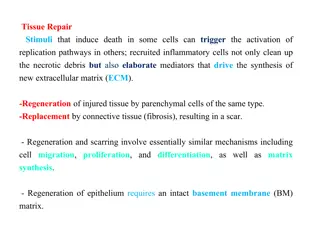
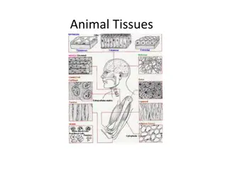
REGENERATION & HEALING BY FIBROSIS Definition of the tissue repair: restoration of tissue architecture and function after an injury. It occurs by two types of reactions: 1-regeneration of the injured tissue . 2- scar formation by the deposition of connective tissue. 1-Regeneration. Some tissues are able to replace the damaged cells and essentially return to a normal state this process is called regeneration. Regeneration occurs by proliferation of residual (uninjured) cells that retain the capacity to divide, and by replacement from tissue stem cells. It is the typical response to injury in the rapidly dividing epithelia of the skin and intestines, and some parenchymal organs, notably the liver. Al-Madena Copy CLS 2
2-Scar formation. 1- If the injured tissues are incapable of regeneration 2- if the supporting structures of the tissue are severely damaged, repair occurs by the laying down of connective (fibrous) tissue, a process that results in scar formation. Although the fibrous scar cannot perform the function of lost parenchymal cells, it provides enough structural stability that the injured tissue is usually able to function. The term fibrosis is most often used to describe the extensive deposition of collagen that occurs in the lungs, liver, kidney, and other organs as a consequence of chronic inflammation, or in the myocardium after extensive ischemic necrosis (infarction).
THE CONTROL OF CELL PROLIFERATION Several cell types proliferate during tissue repair. These include 1. The remnants of the injured tissue (which attempt to restore normal structure) 2. Vascular endothelial cells (to create new vessels that provide the nutrients for the repair process) 3. Fibroblasts (the source of the fibrous tissue that fills defects). Al-Madena Copy CLS 5
4-The proliferation of the above cell types is driven by growth factors. 5-The normal size of cell populations in any given tissue is determined by a balance of cell proliferation, cell death by apoptosis, and emergence of new differentiated cells from stem cells . Al-Madena Copy CLS 6
Mechanisms regulating cell populations. Cell numbers can be altered by increased ordecreased rates of stem cell input, cell death via apoptosis, or changes in the rates of proliferation or differentiation. Al-Madena Copy CLS 7
Proliferative Capacities of Tissues: The tissues of the body are divided into three groups: 1. Continuously Dividing Tissues (labile tissues): cells of these tissues are continuously being lost and replaced by maturation from stem cells and by proliferation of mature cells. Labile cells include: hematopoietic cells in the bone marrow and the majority of surface epithelia. These tissues can readily regenerate after injury provided the pool of stem cells is preserved. Al-Madena Copy CLS 8
2. Stable Tissues: Cells of these tissues are quiescent (in the G0stage of the cell cycle) and have only minimal replicative activity in their normal state. These cells are capable of proliferating in response to injury or loss of tissue mass. Stable cells constitute 1- The parenchyma of most solid tissues, such as liver & kidney. 2- endothelial cells, fibroblasts, and smooth muscle cells; the proliferation of these cells is particularly important in wound healing . With the exception of liver, stable tissues have a limited capacity to regenerate after injury. Al-Madena Copy CLS 9
3. Permanent Tissues: Cells of these tissues are terminally differentiated and nonproliferative in postnatal life. The majority of 1-neurons 2- cardiac muscle cells 3- Skeletal muscle . Accordingly, injury to brain or heart is irreversible and results in a scar. Skeletal muscle is usually classified as a permanent tissue. Al-Madena Copy CLS 10
Stem cells are characterized by two important properties: 1. 1-Self-renewal capacity 2. 2- Asymmetric replication. Asymmetric replication of stem cells means that after each cell division, some progeny enter a differentiation pathway, while others remain undifferentiated, retaining their self- renewal capacity. Al-Madena Copy CLS 11
REPAIR BY CONNECTIVE TISSUE ( scar formation) Healing or repair by connective tissue is encountered if 1. A severe or persistent (chronic) tissue injury that result in damage to parenchymal cells as well as the stromal framework 2. Injury affects non dividing cells Under these conditions, repair occurs by replacement of the non regenerated cells with connective tissue, or by a combination of regeneration of some cells and scar formation.
Repair begins within 24 hours of injury by the emigration of fibroblasts and the induction of fibroblast and endothelial cell proliferation. By 3 to 5 days, a specialized type of tissue that is characteristic of healing, calledgranulation tissue is apparent. The term granulation tissue derives from the pink, soft, granular gross appearance, such as that seen beneath the scab of a skin wound. Its microscopic appearance is characterized by proliferation of fibroblasts and new thin-walled, (angiogenesis), in a loose ECM. delicate capillaries Al-Madena Copy CLS 13
Wound Strength Carefully sutured wounds have approximately 70% of the strength of unwounded skin, largely because of the placement of the sutures. When sutures are removed, usually at 1 week, wound strength is approximately 10% of that of unwounded skin, but this increases rapidly over the next 4 weeks. Collagen synthesis exceeding degradation during the first 2 months, and from structural modifications of collagen (e.g., cross- linking and increased fiber size) when synthesis declines at later times. Wound strength reaches approximately 70% to 80% of normal by 3 months but usually does not substantially improve beyond that point. Al-Madena Copy CLS 14
Causes of delay wound healing 1. Infection is the single most important cause of delay in healing. 2.Nutrition has profound effects on wound healing; protein deficiency & vitamin C deficiency, inhibits collagen synthesis and retards healing. 3. 3. Glucocorticoids (steroids) have anti-inflammatory effects, and their administration may result in poor wound strength due to diminished fibrosis 4.Mechanical variables such as increased local pressureor torsion may cause wounds to pull apart, or dehisce i.e. open out or gape. 5. Poor perfusion, due either to arteriosclerosis and diabetes or to obstructed venous drainage (e.g. in varicose veins), also impairs healing 6. 6. Foreign bodies such as fragments of steel, glass, or even bone impede healing.


")