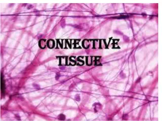
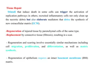
Understanding Repair by Connective Tissue in Healing Processes
Healing or repair by connective tissue occurs in response to severe or chronic tissue injuries, leading to the replacement of nonregenerated cells with connective tissue or scar formation. This process involves the induction of fibroblast and endothelial cell proliferation, granulation tissue formation, and eventual scar development through the sequential processes of angiogenesis, fibroblast migration, ECM deposition, and tissue remodeling. The formation of granulation tissue in wound healing is characterized by proliferation of fibroblasts and capillaries, eventually resulting in scar formation. Cutaneous wound healing exemplifies the principles underlying connective tissue repair in various tissues.

Download Presentation

Please find below an Image/Link to download the presentation.
The content on the website is provided AS IS for your information and personal use only. It may not be sold, licensed, or shared on other websites without obtaining consent from the author. Download presentation by click this link. If you encounter any issues during the download, it is possible that the publisher has removed the file from their server.
E N D
Presentation Transcript
REPAIR BY CONNECTIVE TISSUE DR. AYSER HAMEED LEC.5
Healing or repair by connective tissue is encountered if:- 1. A severe or persistent (chronic) tissue injury that result in damage to parenchymal cells as well as the stromal framework. 2. Injury affects nondividing cells. 2
Under these conditions, repair occurs by replacement of the nonregenerated cells with connective tissue, or by a combination of regeneration of some cells and scar formation. Repair begins within 24 hours of injury by the emigration of fibroblasts and the induction of fibroblast and endothelial cell proliferation. 3
By 3 to 5 days, a specialized type of tissue that is characteristic of healing, called granulation tissue is apparent. The term granulation tissue derives from the pink, soft, granular gross appearance, such as that seen beneath the scab of a skin wound. 4
Its microscopic appearance is characterized by proliferation of fibroblasts and new thin-walled, delicate capillaries (angiogenesis), in a loose ECM. Granulation tissue then progressively accumulates connective tissue matrix, eventually resulting in the formation of a scar, which may remodel over time. 5
Granulation tissue formation in wound healing Rt. There are numerous blood vessels, edema, and a loose extracellular matrix containing occasional inflammatory cells. Lt. at high magnification, granulation tissue has capillaries, fibroblasts, and a variable amount of inflammatory cells. 6
Repair by connective tissue deposition consists of four sequential processes: Formation of new blood vessels (angiogenesis). Migration and proliferation of fibroblasts. Deposition of ECM (scar formation). Maturation and reorganization of the fibrous tissue (remodeling). 7
CUTANEOUS WOUND HEALING This is a process that involves both epithelial regeneration and the formation of connective tissue scar and is thus illustrative of the general principles that apply to wound healing in all tissues. The events are orchestrated by interplay of growth factors and ECM. Cutaneous wound healing has three main phases:- Inflammation. formation of granulation tissue. ECM deposition and remodeling . 8
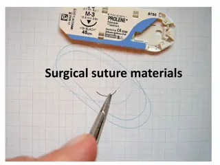
Larger wounds also contract during the healing process. Events in wound healing overlap to a great extent and cannot be completely separated from each other. Based on the nature of the wound, the healing of cutaneous wounds can occur by first or second intention. Healing by First Intention One of the simplest examples of wound repair is the healing of a clean, uninfected surgical incision approximated by surgical sutures. This is referred to as primary union or healing by first intention. 9
The incision causes only focal disruption of epithelial basement membrane continuity and death of a relatively few epithelial and connective tissue cells. As a result, epithelial regeneration predominates over fibrosis. A small scar is formed, but there is minimal wound contraction. The narrow incisional space first fills with fibrin- clotted blood. Within 24 hours, neutrophils are seen at the incision margin, migrating toward the fibrin clot. 10
Within 24 to 48 hours, epithelial cells from both edges have begun to migrate and proliferate along the dermis. The cells meet in the midline beneath the surface scab, yielding a thin but continuous epithelial layer. By day 3, neutrophils have been largely replaced by macrophages, and granulation tissue progressively invades the incision space. Epithelial cell proliferation continues, yielding a thickened epidermal covering layer. By day 5, neovascularization reaches its peak as granulation tissue fills the incisional space. 11
The epidermis recovers its normal thickness as differentiation of surface cells yields a mature epidermal architecture with surface keratinization. During the second week, there is continued collagen accumulation and fibroblast proliferation that bridge the incision. The leukocyte infiltrate, edema, and increased vascularity are diminished. The long process of "blanching" begins, accomplished by increasing collagen deposition within the incisional scar and the regression of vascular channels. 12
By the end of the first month, the scar comprises a cellular connective tissue largely devoid of inflammatory cells and covered by an essentially normal epidermis. The tensile strength of the wound increases with time. However, the dermal appendages destroyed in the line of the incision are permanently lost. 13
Healing by Second Intention (healing by secondary union):- When cell or tissue loss is more extensive, the repair process is more complex, the inflammatory reaction is more intense, there is abundant development of granulation tissue, and the wound contracts by the action of myofibroblasts. 14
This is followed by accumulation of ECM and formation of a large scar. This mode of healing occurs in:- I. Large wounds. II. Abscesses . III.Ulcerations. IV.After infarction in parenchymal organs. 15
Secondary healing differs from primary healing in several respects: A larger clot or scab rich in fibrin and fibronectin forms at the surface of the wound. Inflammation is more intense because large tissue defects have a greater volume of necrotic debris, exudate, and fibrin that must be removed. 16
Much larger amounts of granulation tissue are formed. A greater volume of granulation tissue generally results in a greater mass of scar tissue. 17
Wound healing Steps in wound healing by first intention (left) and second intention (right). In the latter, note the large amount of granulation tissue and wound contraction. 18
The adverse influences on wound healing include: 1. Infection is the single most important cause of delay in healing; it prolongs the inflammation phase of the process and potentially increases the local tissue injury. 2. Nutrition has profound effects on wound healing; protein deficiency & vitamin C deficiency, inhibits collagen synthesis and retards healing. 19
3. Mechanical variables such as increased local pressure or torsion may cause wounds to pull apart, or dehisce i.e. open out or gape. 4. Poor perfusion, due either to arteriosclerosis and diabetes or to obstructed venous drainage (e.g. in varicose veins), also impairs healing 5. Foreign bodies such as fragments of steel, glass, or even bone impede healing. 20
Aberrations of cell growth and ECM production This may occur even in what begins as normal wound healing. 1. Keloid refers to the accumulation of exuberant amounts of collagen that give rise to prominent, raised scars. There appears to be a heritable predisposition to keloid formation, and the condition is more common in blacks. 21
Keloid Excess collagen deposition in the skin forming a raised scar known as a keloid. id. 22
2. Exuberant granulation: healing wounds may also generate excessive granulation tissue that protrudes above the level of the surrounding skin and hinders re-epithelialization. The restoration of epithelial continuity requires cautery or surgical resection of the granulation tissue. 23
3. Disabling fibrosis associated with chronic inflammatory diseases such as rheumatoid arthritis, pulmonary fibrosis, and cirrhosis have many similarities to those involved in normal wound healing. In these diseases, however, persistent stimulation of fibrogenesis results from chronic immune reactions that sustain the synthesis and secretion of growth factors, fibrogenic cytokines, and proteases. 24
Collagen degradation by collagenases, normally important in wound remodeling, is responsible for much of the joint destruction seen in rheumatoid arthritis. 25
Rheumatoid arthritis This deformity of the hand is due to rheumatoid arthritis (RA). This autoimmune disease leads to synovial proliferation and joint destruction, typically in a symmetrical pattern involving small joints of hands and feet, followed by wrists, ankles, elbows, and knees. Rheumatoid factor can be identified serologically in most, but not all, RA patients. 26