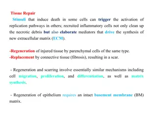
Understanding Glandular Epithelium and Secretory Cells

Glandular epithelium


Glandular Epithelial Tissue
•
Epithelial cells that function mainly to produce
and secrete various macromolecules may
occur in epithelia with other major functions
or comprise specialized organs called
glands.
•
Secretory cells may synthesize, store, and release
proteins (e.g. in the pancreas), lipids (e.g. adrenal,
sebaceous glands), or complexes of carbohydrates
and proteins (e.g. salivary glands). Epithelia of
mammary glands secrete all three substances.
•
The cells of some glands (e.g. sweat glands) have
little synthetic activity and secrete mostly water
and electrolytes (ions) transferred from the blood.
An important, easily seen example is the
goblet
cell
abundant in the lining of the small intestine
•
Types of glands:
1.
Exocrine glands:
remain connected with the surface
epithelium, the connection forming the tubular ducts lined
with epithelium that deliver the secreted material where it is
used (contain duct).
2.
Endocrine glands:
lose the connection to their original
epithelium and therefore lack ducts. Thin-walled blood
vessels (capillaries) adjacent to endocrine cells absorb their
secreted hormone
products for transport in blood to target
cells throughout the
body (without duct).
•
Glands can be
A-
Simple
(ducts not branched) or
B- Compound
(ducts with two or more branches).
■
Secretory portions can be:
•
-
Tubular
(either short or long
and coiled
)
•
-
Acinar
(rounded and saclike); either type
of secretory unit
may be
branched
, even if the duct is not branched.
■
Compound
glands can have branching ducts and can have
multiple tubular, acinar, or tubuloacinar secretory portions.
Three basic mechanisms for releasing the product are
commonly used by cells specialized for secretion, and
cells engaged in each type of secretion can be
distinguished histologically:
1.
Merocrine secretion
:
This is the most common
method of protein or glycoprotein secretion and
involves typical exocytosis from membrane-bound
vesicles or secretory granules.
2.
Holocrine secretion:
Here cells accumulate
product continuously as they enlarge and undergo
terminal differentiation, culminating in complete cell
disruption that releases the product and cell debris
into the gland’s lumen. This is best seen in the
sebaceous glands producing lipid rich material in
skin.
3-
Apocrine secretion:
Here product accumulates
at the cells’ apical ends, portions of which are then
extruded to release the product together with small
amounts of cytoplasm and cell membrane. Lipid
droplets are secreted in the mammary gland in this
manner
Renewal Of Epithelial Cells
•
Epithelial tissues are relatively labile structures
whose cells are renewed continuously by:
-
Mitotic activity
-
Stem cell populations
•
The rate of renewal varies widely; it can be
-
Fast
in tissues such as the intestinal epithelium,
which is replaced every week.
-
Slow
as in the large glands.
•
In stratified epithelial tissues, stem cells and
mitosis occur only within the basal layer in contact
with the basal lamina.
For example, the epithelium
lining the small intestine is derived completely from
stem cells found in the simple glands between the
intestinal villi. In the epidermis, many stem cells are
located at a characteristic position along the wall of
hair follicles
Thank you
Glandular epithelium consists of epithelial cells specialized in producing and secreting various macromolecules, found in glands throughout the body. These secretory cells can synthesize proteins, lipids, and carbohydrates, with different types of glands such as exocrine and endocrine glands. The secretion mechanisms include merocrine and holocrine methods. Understanding the structure and function of glandular epithelium and secretory cells is essential for comprehending gland physiology and pathology.

Download Presentation

Please find below an Image/Link to download the presentation.
The content on the website is provided AS IS for your information and personal use only. It may not be sold, licensed, or shared on other websites without obtaining consent from the author. Download presentation by click this link. If you encounter any issues during the download, it is possible that the publisher has removed the file from their server.
E N D
Presentation Transcript
Glandular Epithelial Tissue Epithelial cells that function mainly to produce and secrete various macromolecules may occur in epithelia with other major functions or comprise specialized organs called glands.
Secretory cells may synthesize, store, and release proteins (e.g. in the pancreas), lipids (e.g. adrenal, sebaceous glands), or complexes of carbohydrates and proteins (e.g. salivary glands). Epithelia of mammary glands secrete all three substances. The cells of some glands (e.g. sweat glands) have little synthetic activity and secrete mostly water and electrolytes (ions) transferred from the blood. An important, easily seen example is the goblet cell abundant in the lining of the small intestine
Types of glands: 1. Exocrine glands: remain connected with the surface epithelium, the connection forming the tubular ducts lined with epithelium that deliver the secreted material where it is used (contain duct). 2. Endocrine glands: lose the connection to their original epithelium and therefore lack ducts. Thin-walled blood vessels (capillaries) adjacent to endocrine cells absorb their secreted hormone products for transport in blood to target cells throughout the body (without duct).
Glands can be A- Simple (ducts not branched) or B- Compound (ducts with two or more branches). Secretory portions can be: - Tubular (either short or long and coiled) - Acinar (rounded and saclike); either type of secretory unit may be branched, even if the duct is not branched. Compound glands can have branching ducts and can have multiple tubular, acinar, or tubuloacinar secretory portions.
Three basic mechanisms for releasing the product are commonly used by cells specialized for secretion, and cells engaged in each type of secretion can be distinguished histologically: 1. Merocrine secretion: This is the most common method of protein or glycoprotein secretion and involves typical exocytosis from membrane-bound vesicles or secretory granules.
2. Holocrine secretion: Here cells accumulate product continuously as they enlarge and undergo terminal differentiation, culminating in complete cell disruption that releases the product and cell debris into the gland s lumen. This is best seen in the sebaceous glands producing lipid rich material in skin.
3- Apocrine secretion: Here product accumulates at the cells apical ends, portions of which are then extruded to release the product together with small amounts of cytoplasm and cell membrane. Lipid droplets are secreted in the mammary gland in this manner
Renewal Of Epithelial Cells Epithelial tissues are relatively labile structures whose cells are renewed continuously by: - Mitotic activity - Stem cell populations The rate of renewal varies widely; it can be - Fast in tissues such as the intestinal epithelium, which is replaced every week. - Slow as in the large glands.
In stratified epithelial tissues, stem cells and mitosis occur only within the basal layer in contact with the basal lamina. For example, the epithelium lining the small intestine is derived completely from stem cells found in the simple glands between the intestinal villi. In the epidermis, many stem cells are located at a characteristic position along the wall of hair follicles