Understanding Cerebrospinal Fluid (CSF): Functions, Formation, and Circulation

Cerebrospinal fluid (CSF) plays a vital role in providing a controlled chemical environment, nutrient supply, waste removal, and physical support and protection to the brain and spinal cord. This fluid circulates around the central nervous system, and its formation involves processes like selective ultrafiltration of plasma and active secretion by epithelial membranes. Understanding the functions, formation, and circulation of CSF is crucial for medical practitioners to diagnose and treat various neurological conditions. Additionally, proper sampling techniques need to be followed to ensure accurate and reliable results in laboratory investigations.

Download Presentation

Please find below an Image/Link to download the presentation.
The content on the website is provided AS IS for your information and personal use only. It may not be sold, licensed, or shared on other websites without obtaining consent from the author. Download presentation by click this link. If you encounter any issues during the download, it is possible that the publisher has removed the file from their server.
E N D
Presentation Transcript
HbA NH2 H2O2 Cl2O7 KClO3 NAOH CH2O PO4 KMnO4 M E D I C I N E COOH KING SAUD UNIVERSITY Co2 MgCl2 H2O SO2 Doctors slides Doctors notes Important ExtraInformation HCN CCl4 CuCl2 SiCl4 Biochemistry Cerebrospinal fluid We can t direct the wind, but we can adjust the sails Editing file
O B J E C T I V E S By the end of this lecture, the students should be able to: Key principles: To identify the CSF functions, formation and circulation. CSF overview Functions, circulation etc. To recognize the method of CSF sampling, and the procedure for specimen collection, and processing CSF investigations and specimen collection To identify the indications and contraindications of lumbar puncture and laboratory investigation of CSF Types and components of CSF examination Electrophoresis To recognize and explain the normal and abnormal findings of physical and biochemical examination of CSF (with special emphasis on the glucose, protein, electrolytes and cellular content of CSF) Abnormal pathological conditions effect To interpret CSF electrophoresis pattern To define expressions describing abnormal locations of CSF as otorrhea and rhinorrhea
CSF Definition & Functions: CSF definition : The liquid surrounding the brain and spinal cord, that flows in subarachnoid space (the area between arachnoid & pia matter) Main Functions: Provides a controlled chemical environment nutrient supply & waste removal Physical support & protection CSF also has a certain transportation functions
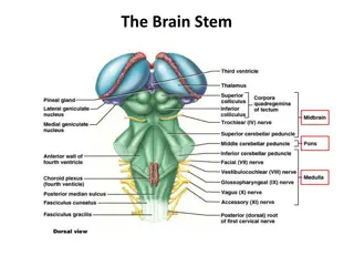
CSF Formation & Circulation: CSF is formed at the choroid plexuses & by the cells lining the ventricles. Normal blood brain barrier is important for the normal chemistry results of CSF Selective ultrafiltration of plasma Active secretion by epithelial membranes Mechanism of formation Rate of formation : Absorption occurs at the arachnoid villi protruding through the dura to the venous sinuses of the brain bloodstream Mechanism of excretion (absorption) Excretion volume = production volume constant CSF volume From CSF to the blood 500 ml/day The rule of equilibrium is production = excretion, but in CSF our absorption is instead excretion
CSF Formation & Circulation: CSF Circulation:
CSF Formation & Circulation: Method of CSF Sampling: The person taking the CSF sample should be professional to prevent traumatic tap (rupturing the blood vessels), because CSF is considered as a precious sample since its difficult to obtain. We always start with the non invasive procedures like taking blood and urine specimens, and then if we needed CSF we may take it. To differentiate between traumatic tap and hemorrhage: 1\ the blood is bright. 2\ the more CSF we withdrawal the lighter the blood becomes because it s a new rupture made to the blood vessel from the needle. Sample is taken by a needle from between the lumbar vertebras L3-L4
CSF Specimen Collection: Obtained bylumbar puncture (At the interspace L3-4, or lower) Using aseptic technique Because CSF has low defense against microbes CSF is separated into 2 aliquots: 1. for chemistry & serology 2. for microbiology and hematology Immediate analysis It s a precious sample: Preserve any remaining sample Extra Extra
CSF investigation Contraindications for performing lumbar puncture: Indications for laboratory investigation of CSF: If the patient has a skin infection at the normal obtaining sites of lumbar puncture what to do ? If the condition is so serious and can not wait then an expert should perform the LP even at the levels of spinal cord above L3 and it has a specific technique .. Otherwise they have to wait until the infection is treated Bleeding diathesis CNS infection Increased intracranial pressure Demyelinating diseases e.g. MS CNS Malignancy Infection at site of needle insertion Hemorrhage
Examination of CSF 1- Physical examination If CSF is cloudy (turbid) Free of blood perform microscopic examination: Normal CSF: Free of clots Colorless Cloudy appearance is usually due to leukocytes Clear And may be due to micro- organisms
IMPORTANT SLIDE Blood & Hemoglobin pigments in CSF: Extra It s important to differentiate between the traumatic and SAH in the blood. When would Xanthochromia indicate hemorrhage? Xantho : Chromia : Hemoglobin has heme (which has a ring that would break down giving us the changing color from red to green) -> like what happens in bruising. Also its not bright, and is color changing If you exclude: Hyperbilirubinemia (bilirubin > 20 mg/dL) Prior traumatic tap A "traumatic tap" occurs if the needle inadvertently has entered an epidural vein during insertion. A yellowish tinge to the CSF fluid is called xanthochromia. Xanthochromia is usually caused by red blood cell degeneration in the CSF as would be seen in subarachnoid hemorrhage (SAH).
Examination of CSF 2- Biochemical analysis of CSF: Glucose Glucose and specific protein tests are the most reliable diagnostically & accessible analytically Specific : 1) Albumin 2) Immunoglobulin 3) Others (e.g. myelin basic protein; MBP) Tests of interest Protein Specimens are sent to the microbiology lab to differentiate whether its bacterial\viral meningitis and glucose\protein are measured in the biochemistry lab. the case will be half micro half biochemistry in the final Total Lactate Lactate not tested any more
CSF Glucose Abnormal CSF glucose IMPORTANT Glucose enters CSF via facilitative transporter (GLUT) Sodium independent CSF [glucose] is ~ 2/3 that of plasma 50 - 80 mg/dl A plasma sample must be obtained ~ 2-4 hours before CSF sample In hypoglycemia: [CSF glucose] may be very low In hyperglycemia: [CSF glucose] is raised. Measure CSF [Glucose]: Immediately Or preserve the specimen with and antiglycolytic Microorganisms eat the glucose and secrete proteins. e.g. fluoride ion Usually they call it a grey tap tube and it has fluoride that would inhibit glycolysis. --> this info is just for your information. A question can be asked: CSF in glucose is more or less than plasma? It is 2/3 glucose of plasma because it enters CSF via facilitative transporter.
CSF proteins Abnormal CSF proteins Proteins, mostly albumin are found in the CSF (0.15-0.45 g/L) We must know how to compare for example if the patient has nephrotic syndrome then his protein levels . Source of CSF proteins: 80% from plasma by ultrafiltration Albumin which is small in size 20% from intrathecal synthesis Immunoglobulin protein which is quite big First we exclude traumatic tap. It will have high protein CSF Albumin Albumin is produced solely in the liver Its presence in CSF must occur through BBB If albumin is normal and IGP is high -> local synthesis disease -> MS (demyelinating disease) SSPE is less common, usually tests in lab are made for MS
CSF proteins What to do if CSF [protein] was detected ? CSF Immunoglobulin Glucose enters CSF via facilitative transporter CSF IgG can arise: 1) Perform electrophoretic separation 2) If multiple banding (oligo-clonal bands) of the -globulin is detected, the following differential diagnosis is suspected: MS SSPE III. Inflammatory diseases a) from plasma cells within CSF b) from the blood through BBB I. II. [IgG] and normal [Alb] of CSF suggests local production of IgG, e.g., Multiple sclerosis (MS) Subacute sclerosing panencephalitis (SSPE)
CSF electrophoresis IMPORTANT SLIDE Oligo-clonal banding MS or SSPE Normal pattern Electrophoresis is a techniques used in labs to separate proteins or immunoglobulins in a specimen (urine blood CSF). Oligo clonal bands are for diagnosing MS Oligo clonal means multiple bands We don t have to know the bands or explain the electrophoresis. We just need to know that oligo clonal gamma means MS and in the normal is a one big spike of monoclonal albumin band and small bands. albumin albumin 1 prealbumin 1 1 2 2 2 prealbumin 1 2
Other Chemical Components of CSF Normal composition of CSF Appearance Clear ,Colorless Lymphocytes <5/mm3 CSF [Calcium], [Potassium] & [Phosphates] are lower than their levels in the blood Polymorphs Nil pH 7.4 CSF [Chloride] & [Magnesium] are higher than their levels in the blood Total Volume 100 - 150 ml Daily Secretion 450 - 500 ml Abnormal CSF [Chloride] Specific Gravity 1.006 - 1.007 marked reduction in acute bacterial meningitis Protein 0.15 0.45 g/L slight reduction in viral meningitis & brain tumors 50 - 80 mg/dL (2.8-4.2 mmol/L) (>50% plasma level) Glucose In TB meningitis chloride and glucose are LOW and protein HIGH. Don t memorize numbers normal ranges are given in test! Chloride 115 - 130 mmol /L Calcium 1.0 - 1.40 mmol/L Phosphorus 0.4 - 0.7 mmol/L Magnesium 1.2 - 1.5 mmol/L Potassium 2.6 - 3.0 mmol/L
Abnormal findings of CSF in some pathological conditions Condition Parameter Bacterial Meningitis (pyogenic) Tuberculous Meningitis Viral Meningitis Appearance Often turbid Often fibrin web Usually clear Predominant cell Polymorphs Mononuclear (lymphocytes) Mononuclear (lymphocytes) Cell count/mm3 90 - 1000+ 10 - 1000 50 - 1000 Bacteria/virus +ve smear & culture Often none in smear negative smear or culture Protein (0.15 - 0.45 g/L) > 1.5 ( ) 1-5 <1 ( ) (Normal) Glucose (2.8 - 4.2 mmol/L) >1/2 plasma (Normal or slightly ) <1/2 plasma ( ) <1/2 plasma ( ) Chlorides (115 - 130 mmol/L) Normal or
Otorrhea & Rhinorrhea Otorrhea : leakage of CSF from the ear Rhinorrhea : leakage of CSF into the nose Extra Extra In Otorrhea blood tests are outdated => MRI is done instead because its faster. Otorrhea happens to people who had a trauma.
Take home messages CSF is formed in the choroid plexus It is essential for the physical protection of the CNS The physical & chemical analysis of CSF is essential for diagnosis of certain diseases Good luck doctors
Summary Function of the CSF (Normal blood brain barrier is important for the normal chemistry results of CSF) 1-Physical support& protection 2-controlled chemical environment . Analysis of CSF Choroid plexuses &ventricle cells (500 ML/day) Physical Formed by Chemical Clear & colorless 1-Selective ultrafiltration of plasma 2-Active secretion by epithelial membranes Mechanism of formation Glucose Protein CSF glu is 2/3 of plasma Albumin & IgG arachnoid villi venous sinuses blood stream Excretion volume = production volume from plasma By GLUT Excretion (absorption) : If IgG is high and albumin in normal : : Total protein : Lumber puncture (L3-L4) Collection of specimen by Hyperglycemia -disorders in carrier mediated trans -Traumatic tap - permeability : Bacterial &fungal infection Cerebral hemorrhage - production by CNS: MS& SSPE -Obstruction : Tumors& abcess MS& SPPE ( meningeal TB & sarcoidosis) *IF oligoclonal bands of Y-globin (MS) 1- CNS infection 2-hemorrhage 3-CNS malignance 4-demylination -active metabolism by microorganism : indications cute purulent, amebic, & fungal meningitis -increased metabolism by cns : 1-Bleeding diathesis 2. intracranial pressure 3. Infection at site of needle Neoplasm Contraindications
Quiz 1) In which of the following situations the lumbar puncture is contraindicated ? 4) What is the situation of CSF glucose level if the patient has bacterial meningitis ? a) b) Multiple sclerosis c) Elevated intracranial pressure d) CNS hemorrhage CNS infection a) b) Decreased c) Depends on the type of the bacteria d) Normal Elevated 2) The appearance of CSF in bacterial meningitis is ? a) b) Fibrin web c) Clear d) None of the above Turbid Q : When would Xanthochromia indicate hemorrhage ? 3) Xanthochromia indicates which of the following ? Q : What is the normal range of CSF proteins ? a) b) Sarcoidosis c) Subarachnoid hemorrhage d) Hyperglycemia Subacute sclerosing panencephalitis (SSPE) 4- B 3- C 2- A 1- C Suggestions and recommendations Suggestions and recommendations
T E A M M E M B E R S TEAM LEADERS Zaina alkaff Mohammad Almutlaq Rania Alessa Haifa alwael
THANK YOU FOR CHECKING OUR WORK Lippincott's Illusrated Reviews Biochemistry 6th E https://www.youtube.com/watch?v=sO1z6Ns3pBg Don t forget to review the notes PLEASE CONTACT US IF YOU HAVE ANY ISSUE @436Biochemteam Biochemistryteam436@gmail.com