Overview of Special Connective Tissue: Cartilage - Histology by Dr. Ammar Ismail

Cartilage is a specialized connective tissue with chondrocytes in lacunae, lacking vasculature and nerves. Hyaline cartilage is common in the body, while elastic cartilage contains elastic fibers, and fibrocartilage is associated with dense connective tissue. Cartilage growth occurs through chondrocyte division and matrix synthesis. Interstitial and appositional growth are two types of cartilage growth.

Download Presentation

Please find below an Image/Link to download the presentation.
The content on the website is provided AS IS for your information and personal use only. It may not be sold, licensed, or shared on other websites without obtaining consent from the author. Download presentation by click this link. If you encounter any issues during the download, it is possible that the publisher has removed the file from their server.
E N D
Presentation Transcript
Histology Special connective tissue By: Dr. Ammar Ismail
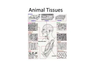
Cartilage: The cartilage possesses cells called chondrocytes which occupy small cavities called lacunae in the extra cellular matrix. The substance of cartilage neither vascularized, nor supplied with nerves or lymphatic vessels. The cells nourishment from blood connective tissues by diffusion through the matrix . The collagen and elastic fibers embedded in the matrix . There are three type of cartilages depended on the fibers which present in the matrix . vessels of surrounded
Hyaline cartilage It is most common cartilage of the body, located in the nose, larynx, tracheal ring, bronchi and in the articular surface of movable joints of the body. This type of cartilage is surrounded by perichondrium which consist of fibroblast and collagen fibers in the outer surface, in the inner surface there are chondroblast which have the ability to produce a new layer of new cartilage .
Histogenesis and growth of cartilage: The un differentiated mesenchymal cells are responsible for hyaline cartilage formation which are aggregation as a dense masses cells which called chondrification differentiate into chondroblasts after losing the cytoplasmic processes become rounded , then turning chondrocytes which have the ability to produce the ground substance and in the same time still capable of cell division to forming a lacuna . centers , these cells
Types of cartilage growth: There are two types of cartilage growth: 1 Interstitial growth: It is occurring only in the early stage also the cartilage grows in the center by chondrocytes division and increase in the ground substance. 2 Appositional growth: In this way the cartilage also grows by adding to its periphery a new cartilage from chondroblast.
Elastic cartilage : The elastic cartilage greatly resembles hyaline except that it is matrix and perichondrium possess elastic fibers, therefore, known as a yellow cartilage. This type of cartilage found in the pinna of ear, epiglottis. cartilage
Fibro cartilage : Is present in the intervertebral discs and in the pubic symphysis, it is associated with hyaline cartilage and with dense connective tissue unlike the other two types of cartilage: 1 fibro cartilage does perichondrium therefore the growth occur by differentiation of fibroblast connective tissue which found aside to the chondrocytes. not possess a in the dense 2 The ground substance is very few because of there are a large amount of collagenous bundle and chondrocytes. 3 The cells are small in the lacuna and appear as a short strip .
Bone : The bone is a specialized connective tissue consists of cells, substance. Whose extra cellular matrix is calcified, the bone is nourishment by blood vessels. The bone characterized appositional growth system. fibers and ground by has haversian only and have
Bone functions : 1 The bone tissue form a skeleton of body for support and protection of the soft organs of the body including brain , spinal cord , heart , lungs . 2 Supporting structure for muscles. 3 Consider as a source for several minerals of the body (store about 99% of the body's calcium). 4 The bone contains the marrow cavity which houses the bone marrow (a hemopoietic organ).
Types of bones: The gross observation of bone shows two types of bones: 1 Compact bone (cortical bone ) : It s a hard mass of bone substance. The bone is covered on its external surface, except at synovial articulations, with periosteum which consist of an outer layer of dense fibrenous connective tissue and an inner cellular osteogenic cells, also the central cavity of bone is lined by endosteum. 2 Spongy bone (cancellous bone ) (trabecular bone): Consist of irregular interconnecting bars , the trabeculae , forming three dimensional net work of lined spaces filled with bone marrow .
Cells of the bone 1 - Osteoprogenitor cells : The cells are located in the inner cellular layer of the periosteum endosteum, these cells derived from the mesenchymal cells. they have ability to differentiated to the osteoblast. . and in the
2 Osteoblast : Derived from osteoprogenitor cells, are responsible for the synthesis of organic components of the bone matrix including collagen and ground substance. Its located on the surface of the bone.
3 Osteocytes Are mature bone cells, derived from osteoblasts. They are housed in the lacunae within the calcified bone matrix.
4 osteoclasts They are large multinucleated cells, derived from the fusion of (monocytes). They are occupies called Howship,s lacunae on the surface of the bone. play an important role of bone resorption, these mechanism hormones ,parathyroid and calcitonin. many blood cells shallow depressions controlled by two
Bone Matrix : The bone matrix is consisting of collagen fibers embedded in substance. The hardness substance due to which form 65% while organic components form 35 % , often the inorganic material is formed from crystal of calcium phosphate . The bone matrix is precipitate as a lamellae and the fibers in each lamella are parallel the hard of components ground ground inorganic
Bone structure: The structural unit of the compact bone is a haversian system system is composed of cylinders of lamellae , concentrically arranged around a vascular space known as the haversian canal .each lamella consist of oval small osteocyte which located in the lacuna and from the lacuna extend a small canaliculi which occupies by a cytoplasmic osteocyte and by which the communication occur among osteocytes osteocytes and haversian canal . Haversian canal of the adjacent connected to each other by transversal canal called Volkmann,s canals . The interstitial space between Osteons is filled by bone lamellae called interstitial lamellae , the outer circumferential lamellae are just deep to the attached to the periosteum by sharpies fibers , the inner circumferential lamellae just deep to the endosteum ( Osteons ) , each process of and between Osteons are periosteum and
Histogenesis and growth of bone: The bone formation during embryonic development (fetal period) occur by two types, intramembranous and endochondreal. The growth of bone occurs only by Appositional growth and because of the hard ground substance the interstitial growth not occurs.
Intra membranous bone formation Most flat bone ( scapula, frontal, ramus, clavical bones formed by intra membranous formation . The undifferentiated mesenchymal cells differentiate into osteoblasts that secrete bone matrix forming network of specules and trabiculae , this region of initial osteogenesis is known as the primary ossification center .Calcification occur and the osteoblasts trapped in their matrix become osteocytes . The mesenchymal cells provide a supply of undifferentiated osteoprogenitor cells which form osteoblasts. As the sponge like network of trabeculae is established, the vascular connective transformed into bone marrow and forming spongy bone and when added additional bone lamellae the spongy bone transferred to the compact bone. tissue in there interstitial
Endochondreal bone formation: Most endochonderial bone formation. The hyaline cartilage model is formed at first then the cells in the center of cartilage are hypertrophy and mature then the matrix among the cells become little after that the chondrocytes begin release the alkaline phosphatase which play role in digestion the cartilage matrix after that, the capillaries in the perichondrium invaded the matrix then the matrix calcified and the chondrocytes will die in the same time the undifferentiated mesenchymal cells in perichondrium differentiated to Osteoblast , then this cells begin to produce bone matrix . The perichondrium become periosteum , the osteoclasts remove mineralized osseous matrix in the center and gradually the center of bone is empty to occupies by marrow . of long and short bones of body developed by