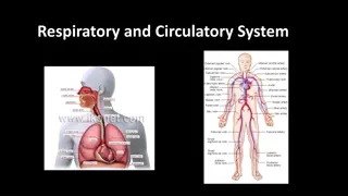
Development of the Respiratory System in Human Embryos
The formation of the lung buds, larynx, trachea, bronchi, and lungs in human embryos involves complex processes where various structures develop from different tissue origins. The lung buds arise from the ventral wall of the foregut, while the larynx, trachea, and bronchi epithelium are of endodermal origin. Mesoderm contributes to the cartilaginous and connective tissue components. The larynx undergoes morphological changes, and the trachea, bronchi, and lungs differentiate during the fifth week of development.

Download Presentation

Please find below an Image/Link to download the presentation.
The content on the website is provided AS IS for your information and personal use only. It may not be sold, licensed, or shared on other websites without obtaining consent from the author. Download presentation by click this link. If you encounter any issues during the download, it is possible that the publisher has removed the file from their server.
E N D
Presentation Transcript
Respiratory System FORMATION OF THE LUNG BUDS When the embryo is approximately 4 weeks old, the respiratory diverticulum (lung bud) appears as an outgrowth from the ventral wall of the foregut ,epithelium of the internal lining of the larynx, trachea, and bronchi, as well as that of the lungs, is entirely of endodermal origin. The cartilaginous, connective tissue components of the trachea and lungs are derived mesoderm surrounding the foregut. muscular, and from splanchnic
Initially, the lung bud is in open communication with the foregut (Fig. diverticulum expands caudally, however, two longitudinal ridges, the tracheoesophageal ridges, separate it from the Subsequently, when these ridges fuse to form the tracheoesophageal septum, the foregut is divided into a dorsal portion, the esophagus, and a ventral portion, the trachea and lung buds (Fig. 13.2B,C). The respiratory primordium communication with the pharynx through the laryngeal orifice (Fig. 13.2D). 13.1B). When the foregut (Fig. 13.2A). maintains its
LARYNX The internal lining of the larynx originates from endoderm, but the cartilages and muscles originate from mesenchyme of the fourth and sixth pharyngeal arches. As a result of rapid proliferation of this mesenchyme, the laryngeal orifice changes in appearance from a sagittal slit to a T-shaped opening (Fig. 13.4A). Subsequently, when mesenchyme of the two arches transforms into the thyroid, cricoid, and arytenoid cartilages, the characteristic adult shape of the laryngeal orifice can be recognized
At about the time that the cartilages are formed, the laryngeal epithelium also proliferates rapidly, resulting in a temporary occlusion of the lumen. Subsequently, vacuolization and recanalization produce a pair of lateral recesses, the laryngeal ventricles. These recesses are bounded by folds of tissue that differentiate into the false and true vocal cords. Since musculature of the larynx is derived from mesenchyme of the fourth and sixth pharyngeal arches, all laryngeal muscles are innervated by branches of the tenth cranial nerve, the vagus nerve. The superior laryngeal nerve innervates derivatives of the fourth pharyngeal arch, and the recurrent laryngeal nerve innervates derivatives of the sixth pharyngeal arch
TRACHEA, BRONCHI, AND LUNGS During its separation from the foregut, the lung bud forms the trachea and two lateral outpocketings, buds . At the beginning of the fifth week, each of these buds enlarges to form right and left main bronchi. The right then forms bronchi, and the left, two , thus foreshadowing the three lobes on the right side and two on the left . the bronchial three secondary
With subsequent growth in caudal and lateral directions, the lung buds expand into the body cavity . The spaces pericardioperitoneal canals, are narrow. They lie on each side of the foregut and are gradually filled by the expanding lung buds. Ultimately the pleuroperitoneal and separate the pericardioperitoneal canals from the peritoneal and pericardial cavities, respectively, and the remaining spaces form the primitive pleural cavities. The mesoderm, which covers the outside of the lung, develops into the visceral pleura. The somatic mesoderm layer, covering the body wall from the inside, becomes the parietal pleura . The space between the parietal and visceral pleura is the pleural cavity for the lungs, the pleuropericardial folds
During further development, secondary bronchi divide repeatedly in a dichotomous fashion, forming ten tertiary (segmental) bronchi in the P.204 right lung and eight in the left, creating the bronchopulmonary segments of the adult lung. By the end of the sixth month, approximately 17 generations of subdivisions have formed. Before the bronchial tree reaches its final shape, however, an additional six divisions form during postnatal life. Branching is regulated by epithelial-mesenchymal interactions between the endoderm of the lung buds and splanchnic mesoderm that surrounds them. Signals for branching, which emit from the mesoderm, involve members of the fibroblast growth factor family. While all of these new subdivisions are occurring and the bronchial tree is developing, the lungs assume a more caudal position, so that by the time of birth, the bifurcation of the trachea is opposite the fourth thoracic vertebra.
MATURATION OF THE LUNGS (TABLE 13.1) Up to the seventh bronchioles divide continuously into more and smaller canals (canalicular phase) (Fig. 13.8A), and the vascular supply increases steadily. Respiration becomes possible when some of the cells of the cuboidal respiratory bronchioles change into thin, flat cells (Fig. 13.8B). These cells are intimately associated with numerous blood and lymph capillaries, and the surrounding spaces are now known as terminal sacs or primitive alveoli. During the seventh month, sufficient numbers of capillaries are present to guarantee adequate gas exchange, and the premature infant is able to survive prenatal month, the
During the last 2 months of prenatal life and for several years thereafter, the number of terminal sacs increases steadily. In addition, cells lining the sacs, known as type I alveolar epithelial cells, become thinner, so that surrounding capillaries protrude into the alveolar sacs (Fig. 13.9). This intimate contact between endothelial cells makes up the blood-air barrier. Mature alveoli are not present before birth. In addition to endothelial cells and flat alveolar epithelial cells, another cell type develops at the end of the sixth month. These cells, type II alveolar epithelial cells, produce surfactant, a phospholipid-rich fluid capable of lowering surface tension at the air-alveolar interface epithelial and
The amount of surfactant in the fluid increases, particularly during the last 2 weeks before birth. As concentrations of surfactant increase during the 34th week of gestation, Fetal breathing movements begin before birth and cause aspiration of amniotic fluid. These movements are important for stimulating lung development and conditioning muscles. When respiration begins at birth, most of the lung fluid is rapidly resorbed by the blood and lymph capillaries, and a small amount is probably expelled via the trachea and bronchi during delivery. respiratory
Respiratory movements after birth bring air into the lungs, which expand and fill the pleural cavity. Although the alveoli increase somewhat in size, growth of the lungs after birth is due primarily to an increase in the number of respiratory bronchioles and alveoli. It is estimated that only one-sixth of the adult number of alveoli are present at birth. The remaining alveoli are formed during the first 10 years of postnatal life through the continuous formation of new primitive alveoli.


")