Overview of Mammalian Respiratory System Histology
The respiratory system in mammals functions to warm, humidify, and cleanse the air as it travels from the nostrils to the lungs. It comprises the nasal cavity, pharynx, larynx, trachea, bronchi, and bronchial tree. The epithelial linings vary in different parts, containing specialized cells and glands. The larynx is supported by elastic and hyaline cartilage, with skeletal muscles contributing to its structure.

Download Presentation

Please find below an Image/Link to download the presentation.
The content on the website is provided AS IS for your information and personal use only. It may not be sold, licensed, or shared on other websites without obtaining consent from the author. Download presentation by click this link. If you encounter any issues during the download, it is possible that the publisher has removed the file from their server.
E N D
Presentation Transcript
Histology Respiratory System
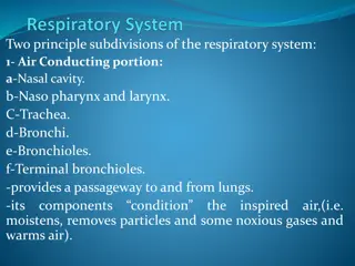
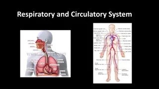
Respiratory system mammals Air flows from the nostrils through a system of passages to the respiratory surfaces of the lungs. As it progresses, it becomes warmed, humidified, and cleansed of some of its particulate matter. Dust, which finds its way to the alveoli, is ultimately consumed by macrophages patrolling the tiny cul-de-sacs. The major components of the air-passage system are the nasal cavity, pharynx, larynx, trachea, bronchi, and the various smaller subdivisions of the bronchial tree leading to the alveoli .
Air from the naris enters the vestibule, the first part of the nasal cavity. The vestibule is lined by a stratified squamous epithelium, which is continuous with the skin externally and with the respiratory portion of the nasal cavity internally. In the horse hairy skin continues into the vestibule. A lamina propria and under lying submucosa support epithelium. the vestibular
The respiratory portion of the nasal cavity is lined pseudostratified epithelium with goblet cells. The lamina propria tubuloalveolar glands. The latter are mainly serous, but mucous and mixed glands do occur. Glands are sparse in carnivores. A submucosa supports the lamina propria. by a ciliated, columnar contains
The (pseudostratified composed of olfactory (sensory) cells, supporting cells, and basal cells. Bowman's glands, tubular and mucoserous, occur within the lamina propria. They open to the surface through ducts lined by cuboidal or flattened submucosa lies below the lamina propria. olfactory epithelium columnar) is cells. A
The nasopharynx and oropharynx are subdivisions of the pharynx. The former is lined by , pseudostratified columnar epithelium with goblet cells, whereas the latter is covered by a stratified squamous epithelium. The lamina propria contains tubular mixed glands in the nasopharynx and mucous glands in the oropharynx.
The larynx is lined in part by a stratified squamous epithelium and partly by a ciliated, pseudostratified columnar epithelium. Numerous elastic fibers are present in the lamina propria. Glands (serous, mucous, and mixed) occur in the lamina propria and submucosa, but are lacking in the vocal and vestibular folds. Hyaline and elastic cartilage provide support for the laryngeal wall. The elastic cartilage of the epiglottis may be partially or completely replaced by adipose tissue, as in carnivores. Skeletal muscles are an integral part of the laryngeal structure
Trachea The trachea is lined by a ciliated, pseudostratified columnar with goblet cells. A lamina propria and submucosa lie below the epithelium, but are not clearly demarcated from one another. Glands, mostly mixed, occur in the deeper layers of the lamina propria and within the submucosa. Rings of hyaline cartilage, which are incomplete dorsally, support the tracheal wall. A layer of smooth muscle, the trachealis muscle, is located trachea. It is positioned internal to the gap in the tracheal cartilages in the horse, pig, and ruminants. in the cat and dog it lies external to the gap. An adventitia of connective tissue completes the wall of the trachea. epithelium dorsally in the
bronchi A ciliated, pseudostratified columnar epithelium with goblet cells lines the bronchi. The epithelium becomes reduced in height as the caliber of the bronchi diminishes. The lamina propria is surrounded by a layer of obliquely arranged smooth muscle. The connective tissue external to the musculature contains mixed glands and plates of hyaline cartilage. In the cat the bronchial cartilages may contain elastic fibers. When seen in histologic sections, the mucosa of large bronchi has few folds. Folds increase as the bronchi decrease in diameter.
The smallest bronchi give rise to suborders of bronchioles. The smallest of the latter, the terminal bronchilioles, branch into two or more respiratory bronchioles, which divide into alveolar ducts that, in turn, empty into alveolar sacs. Bronchioles lack cartilage and glands. Glands, however. Spirally or obliquely arranged smooth muscle forms part of the wall of a bronchiole. The amount of smooth muscle is proportional to the size of the bronchiole. Large bronchioles are lined by ciliated columnar cells, whereas the smallest (terminal) bronchioles are lined proximally by ciliated cuboidal cells and, distally, by non ciliated cells. The mucosa of the bronchioles is folded.
Respiratory bronchioles branch from the ends of terminal bronchioles. They are lined by a cuboidal epithelium, which becomes flattened distally, and their wall contains some smooth muscle. Alveoli are scattered within the epithelium. Respiratory bronchioles are best developed in the cat and dog.
Alveolar ducts branch from respiratory bronchioles. Their thin walls are constructed entirely of alveoli. The lip of each alveolus of an alveolar duct contains smooth muscle arranged circumferentially. The presence of the muscle gives the lip of the alveolus a knoblike appearance when histologic sections occur at right angles to the long axis of the muscle cells. Ultimately, each alveolar duct branches into three or more alveolar sacs. No smooth muscle is present in the sacs. Therefore, the alveoli, which form the walls of the sacs, do not have lips with knoblike expansions as do those of the alveolar ducts. Alveoli are lined mainly by exceedingly thin squamo us epithelial cell s (type I cell s). Alveoli are separated from one another by a thin, highly vascularized layer of fine collagenous and elastic fibers. This layer, together with the squamous cells lining the adjacent alveol i, forms an alveolar septum.