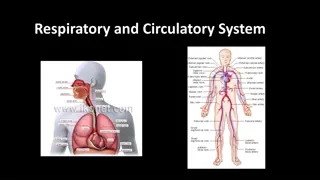
Overview of the Respiratory System

The human respiratory system facilitates gas exchange crucial for cellular energy production. Comprised of the upper and lower respiratory systems, it filters, warms, and humidifies air before reaching the alveoli for efficient exchange. Functions include gas exchange, air movement, and protection from pathogens.

Download Presentation

Please find below an Image/Link to download the presentation.
The content on the website is provided AS IS for your information and personal use only. It may not be sold, licensed, or shared on other websites without obtaining consent from the author. Download presentation by click this link. If you encounter any issues during the download, it is possible that the publisher has removed the file from their server.
E N D
Presentation Transcript
The Respiratory System (or Pulmonary System): Living cells need energy for maintenance, growth, defense & replication. Human cells obtain that energy through aerobic mechanisms that require oxygen & produce carbon dioxide. Humans respiratory exchange surfaces are confined to the inside of the lungs (in environment) under these conditions, diffusion can occur between the air and blood. The cardiovascular system provides a link between the interstitial fluids & the exchange surfaces of the lungs. (the circulating blood carries oxygen from the lungs to peripheral tissue, it also accepts & transports carbon dioxide generated by those tissues delivering it to the lungs. a warm, moist, protected
Diffusion between the blood & air occurs at the alveoli of the lungs. The distance between the blood in an alveolar capillary & the air inside an alveolus is generally less than 1 m & in some cases as small as 0.1 m. the total surface area for gas exchange in adult lungs is 70m to 140m . Functions of the respiratory system: 1.providing an extensive area for gas exchange between the air & circulating blood. 2.moving air to and from the exchange surface of the lung. 3.protecting respiratory surfaces temperature changes & defending the respiratory system & other tissue from invasion by pathogens. 4.Producing sounds involved communication. 5.providing olfactory sensation to the CNS from the olfactory epithelium in the nasal cavity. from dehydration, in speaking &
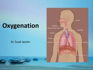
Organization of the respiratory system: Upper respiratory system = consist of the nose, nasal cavity, Para nasal sinuses & Pharynx. (these passageways filter, warm & humidify the incoming air (protecting the more delicate surfaces of the lower respiratory sys.) cooling & dehumidifying outgoing air). Lower respiratory system = include the larynx (voice box), trachea, bronchi, bronchioles & alveoli of the lungs. The respiratory tract consists of the airways that carry air to & from the exchange surface of the lung , so the respiratory tract can be divided to ( Conducting portion & Respiratory portion ).
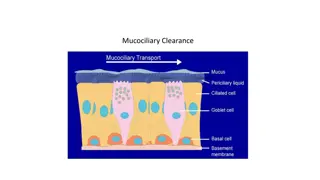
-Conducting portion = begins at the entrance to the nasal cavity & extends through the pharynx & larynx & along the trachea , bronchi & bronchioles to the terminal bronchioles . -Respiratory portion = includes the delicate respiratory bronchioles & the sites of gas exchange (alveoli ). So the filtering , warming & humidification of the inspired air is at the entrance to the upper respiratory system & continue throughout the rest of the conducting system . When the air reaches the alveoli , most foreign particles & pathogens are removed & the humidity & temperature are within acceptable limits this is due to properties of respiratory mucosa .
The Respiratory Mucosa :- Respiratory mucosa lines the conducting portion of the respiratory system & the mucosa is a mucous membrane consists of an epithelium & an underlying connective tissue . Respiratory Epithelium = a pseudo stratified , ciliated , columnar epithelium with numerous goblet cells. Lines the nasal cavity & the superior portion of the pharynx . The structure of the respiratory epithelium change as you proceed along the respiratory tract . The epithelium lining the inferior portions of pharynx is a stratified squamous epithelium similar to that of oral cavity (these portions of the pharynx which conduct air to the lower respiratory tract also conduct food to the esophagus ) . At the beginning of the lower respiratory tract is a pseudo stratified ciliated columnar epithelium comparable to that of nasal cavity .
In the smaller bronchioles , these pseudo stratified epithelium is replaced by a cuboidal epithelium with few cilia . The exchange surface of the alveoli are lined by a very delicate simple squamous epithelium . Lamina Propria = is the underling layer of loose connective tissue that supports the respiratory epithelium . In the upper respiratory system & in the trachea & bronchi , the lamina propria contain mucous glands ( that discharge there secretions onto the epithelial surface . In the conducting portions of lower respiratory system contains bandles of smooth muscle cells .
The Upper Respiratory System :- Nose , Nasal Cavity , Para nasal sinuses & Pharynx . The nose: is the primary passage way for air entering the respiratory system , air normally enters the respiratory system through paired (external nares ) or nostrils which open into the nasal cavity . The vestibule is the space contained within the flexible tissue of the nose , the epithelium of the vestibule contains coarse hairs that extend across the external nares (large air borne particles , such as sand , sawdust , insects are trapped in these hairs & therefore prevented from entering the nasal cavity ). The mucosa of nasal cavity prepares the air for the lower respiratory , it has extensive vascularization & this provides a mechanism of warming & humidifying the incoming air ( as well as for cooling & dehumidifying out going air ) .
As cool , dry air passes inward over the surface of the nasal cavity , the warm epithelium radiates heat & the water in the mucous evaporates , air moving from the nasal cavity to the lungs has been heated almost to body temperature & nearly saturated with water vapor . So breathing through the mouth eliminates much of the filtration , heating &humidifying of inspired air . The pharynx: is a chamber shared by the digestive & respiratory system , it extends between the internal nares & the entrance to the larynx & esophagus , its divided into three regions :-
a- nasopharynx is the superior portion of the pharynx , its connected to the posterior portion of the nasal cavity through the internal nares &is separated from the oral cavity by the soft palate , the nasopharynx is lined by the same pseudo stratified ciliated columnar epithelium as that of the nasal cavity. b- oropharynx extends between the soft palate & the base of the tongue. The posterior portion of the oral cavity communicates directly with the oropharynx & the epithelium changes from a pseudo stratified columnar epithelium to a stratified squamous epithelium . c- laryngopharynx is the inferior portion of pharynx lies between the base of the tongue & the entrance to the larynx & esophagus & like the oropharynx , its lined by a stratified squamous epithelium ( that can resist mechanical abrasion , chemical attack & pathogenic invitation .
The Lower Respiratory System :- Larynx , Trachea , Bronchi , Bronchioles & Alveoli of lungs . The Larynx :- The larynx begins at the level of vertebra C4 or C5 & ends at the level of C6 & it s a cylinder , incomplete cartilaginous walls and stabilized by ligaments & skeletal muscles . Air passing through the glottis vibrates the vocal fold & produce sound waves, the pitch of the sound produced depends on the diameter, length & tension in the vocal folds (diameter & length are related to the size of the larynx).
The Trachea :- is a tough, flexible tube with a diameter of about 2.5cm(1in) approximately 11cm(4.25in). the mucosa of the trachea resembles that of the nasal cavity & nasopharynx. The submucosa is a thick layer of connective tissue that surrounds the mucosa. & length of The trachea contains 15-20 tracheal cartilages these bound together by elastic annular ligaments, the cartilages stiffen the tracheal walls & prevents its collapse or over-expansion.
Each tracheal cartilage is C-shaped, the closed portion of the C protects the anterior & lateral surfaces of the trachea, the open portion of the C faces posteriorly towards the esophagus (the posterior tracheal wall can distort when we swallow, so large masses of food can pass through the esophagous). The ends of each tracheal cartilage are connected by a band of smooth muscle called the Trachealis, contraction of the trachealis alters the diameter of the trachea changing the resistance to air flow.
The Primary Bronchi :- the trachea branches within the mediastinum to right & left primary bronchi supplying the right & left lungs. Primary bronchi is the same as the trachea in histological organization. The Lungs :- the left & right lungs are situated in the left & right plural cavity, with the apex superior to the first rib & the base of each lung rests on the diaphragm. - right lung has three lobes (superior, middle, inferior). - left lung has two lobes (superior, inferior).
The Bronchi :- the primary bronchi & their branches form the Bronchial tree. Each primary bronchus divides to form secondary bronchi (or lobar bronchi), the right primary bronchus divide into three secondary bronchi (superior, middle & inferior lobar bronchi) & the left primary bronchus divide into two secondary bronchi (superior & inferior lobar bronchi). Within each lung the secondary bronchi branch to form tertiary bronchi (or segmental bronchi). The walls of the secondary & tertiary bronchi contain progressively lesser amount of cartilages, and as the amount of cartilages decrease the amount of smooth muscles increase (so the amount of tension in the smooth muscles has great effect on bronchial diameter & great effect on the resistance to air flow.
The Bronchioles :- each tertiary bronchus branches several times to give rise to multiple bronchioles, these branches further into finest conducting branches called terminal bronchioles about 6500 terminal bronchioles are supplied by each tertiary bronchus. Walls of the bronchioles has no cartilages so they are formed by smooth muscles, varying the diameter of the bronchioles by the smooth muscles provides control over the amount of resistance to airflow & the distribution of air within the lungs. The nervous system regulates the activity of this smooth muscle layer & therefor controls the diameter of bronchioles. Sympathetic activity leads to enlargement of airway diameter or bronchodialation. parasympathetic stimulation leads to bronchoconstriction which is a reduction in diameter of airways.
Terminal bronchioles branches to form several respiratory bronchioles & these are the thinnest & most delicate branches of the bronchial tree, they deliver air to the exchange surface of the lung. The Alveoli :- the respiratory bronchioles are connected to multiple alveoli along regions called alveolar ducts, these passageways end at alveolar sacs. Each lung contains about 150 million alveoli, each alveolus has an extensive network of capillaries which are surrounded by a network of elastic fibers. The Alveolus has three types of cells: alveolar epithelium consists of simple squamous epithelium (or type I cells) they are thin and delicate. alveolar macrophages (or dust cells).
squamous cells & they produce an oily secretion (or surfactant) which forms a superficial coating over the thin layer of water in the alveoli. Surfactant reduces the surface tension in the liquid coating the alveolar surface (the surface tension result from attraction between water molecules), so it protects the alveoli from collapsing. surfactant cells (or type II cells) are between the
The Respiratory Membrane: consisting of three parts= 1. the squamous epithelial cells lining the alveolus. 2. the endothelial cells lining the capillary. 3. the basement membrane that lie between the alveolar & endothelial cells. *diffusion across the respiratory membrane proceeds very rapidly because: - the distance is small between the alveolar air & blood. - both O2 & Co2 are lipid soluble.
:")