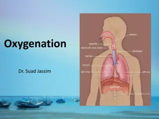
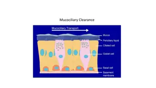
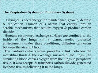
Methods of Imaging the Respiratory System
This lecture discusses various methods of imaging the respiratory system, including plain films, radionuclide imaging, CT scans, ultrasound for pleural disease, MRI, and bronchography. The normal anatomy relevant to respiratory system angiography is also highlighted through a series of images.

Download Presentation

Please find below an Image/Link to download the presentation.
The content on the website is provided AS IS for your information and personal use only. It may not be sold, licensed, or shared on other websites without obtaining consent from the author. Download presentation by click this link. If you encounter any issues during the download, it is possible that the publisher has removed the file from their server.
E N D
Presentation Transcript
Methods of imaging the respiratory system 1. Plain films 2. Radionuclide imaging 3. CT 4. US (for pleural disease) 5. MRI 6. Bronchography.
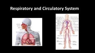
The normal anatomy necessary to the respiratory system angiography
The normal anatomy necessary to the respiratory system angiography
The normal anatomy necessary to the respiratory system angiography
The normal anatomy necessary to the respiratory system angiography
The normal anatomy necessary to the respiratory system angiography
The normal anatomy necessary to the respiratory system angiography
The normal anatomy necessary to the respiratory system angiography
The normal anatomy necessary to the respiratory system angiography
The normal anatomy necessary to the respiratory system angiography
The normal anatomy necessary to the respiratory system angiography
The normal anatomy necessary to the respiratory system angiography
The normal anatomy necessary to the respiratory system angiography
The normal anatomy necessary to the respiratory system angiography
The normal anatomy necessary to the respiratory system angiography
Abnormal Pulmonary angiography pulmonary embolism (PE)
Abnormal Pulmonary angiography pulmonary embolism (PE)
Abnormal Pulmonary angiography pulmonary embolism (PE)
Pulmonary arteriography is the gold standard and will detect most pulmonary emboli. Technique 1. Unenhanced scan of thorax - to detect any other clinical abnormality which might account for symptoms. 2. Enhanced scan of pulmonary arterial system
Patient preparation Prepare The D-dimer blood test (norm<500) Blood urea + serum creatinine patient position supine with their arms above their head scan extent lunge apices to diaphragm Remove metals attached to the clothes Blue canula or pink , test the canula ,
During exam Use manual method (look at the superior vena cava) Respiration phase inspiration monitoring slice (region of interest) below the carina ( Trachea bifurcation ) at the level of the pulmonary trunk. Use bolus method A-IF ROI on the SVC the HU would be approx:175 HU B- IF the ROI on the pulmonary trunk ,The HU would be 100 HU Contrast injection (1or 2 ml per kg ), (usually 75-85 ml) **In the perfect scan, the contrast should not appear in the descending aorta. ** Before the radiation starts make sure the CT scan room door is closed **During IV administration Look at the patient and your CT screen to make sure that the cannula and the connection line is working properly during the exam
The normal anatomy necessary to the respiratory system angiography Topogram
A GUIDE TO CT pulmonary angiogram (CTPA) procedure (manual method) Volume of contrast medium - 80 ml Contrast (and 20 ml of normal saline). Delay usually around 15 s (Manual look at the pulmonary trunk or the SVC when the contrast reach press start scanning) Rate of injection - 4 ml/s Collimation - 3 mm Pitch - 1.8 Start point - just above the aortic arch End point - the lowest hemidiaphragm.
A GUIDE TO CT pulmonary angiogram (CTPA) procedure (Auto method ) Volume of contrast medium 80 ml Contrast +20 ml Normal saline ROI circle - If on the pulmonary trunk HU = 100 - If on the SVC HU = 175 Rate of injection 4 - 6 ml/s Start point - just above the aortic arch End point - the lowest hemidiaphragm.
Practical test of the exam Practical CTPA , Toshipa (Canon) 64 slice CT scan Please Watch carefully !!
References Further reading Klein, J.S. (ed) (2000) Interventional Chest Radiology. Radiol. Clin. N. Am. 38 (2). Macklem P.T. (1998) New methods of imaging the respiratory system. Respirology 3, 101-102. PIOPED Investigators. (1990) Value of ventilation/perfusion scanning in acute pulmonary embolism. JAMA 263 , 2753-2759. Robinson, P.J. (1989) Lung scintigraphy. Clin. Radiol. 4 0 , 557-560. Further reading Remy-Jardin, M., Artaud, D., Fribourg, M. & Beragi, J.P. (i 998) Spiral CT of pulmonary emboli: diagnostic approach, interpretative pitfalls and current indications. Eur. Radiol. 8, 1376-1390.


")
")