Understanding the Respiratory System and Its Functions


Respiratory
Physiology
Dr. Aida Korish
Associate Prof.
Physiology
KSU
Dr.Aida
Korish (
akorish@ksu.edu.sa)
The main goal
of
respiration is
to
1
Provide
oxygen
to
tissues
2
Remove
CO2
Respiratory system consists
of:
•
passages
(airways)
•
muscles
•
centers
Dr.Aida
Korish (
akorish@ksu.edu.sa)
Dr.Aida
Korish (
akorish@ksu.edu.sa)
F
u
n
c
t
i
o
n
s
a
n
d
o
r
g
a
n
i
z
a
t
i
o
n
o
f
t
h
e
r
e
s
p
i
r
a
t
o
r
y
s
y
s
t
e
m
Learning
Objectives
•
By
the
end
of
this
lecture
you will
be able
to:-‐
Dr.Aida
Korish (
akorish@ksu.edu.sa)
1-
De
sc
rib
e
th
e
st
ructu
r
e
s
an
d
func
ti
ons
o
f
the
conductive
and respiratory
zones of
airways.
2-‐Understand
the difference
between
internal and
external
respiration.
3-‐Understand
the
functions of
the respiratory
system,
including
non-‐respiratory
functions,
like clearance
mechanism
by
mucus
and
cilia, production
of
surfactant and its
physiological
significance.
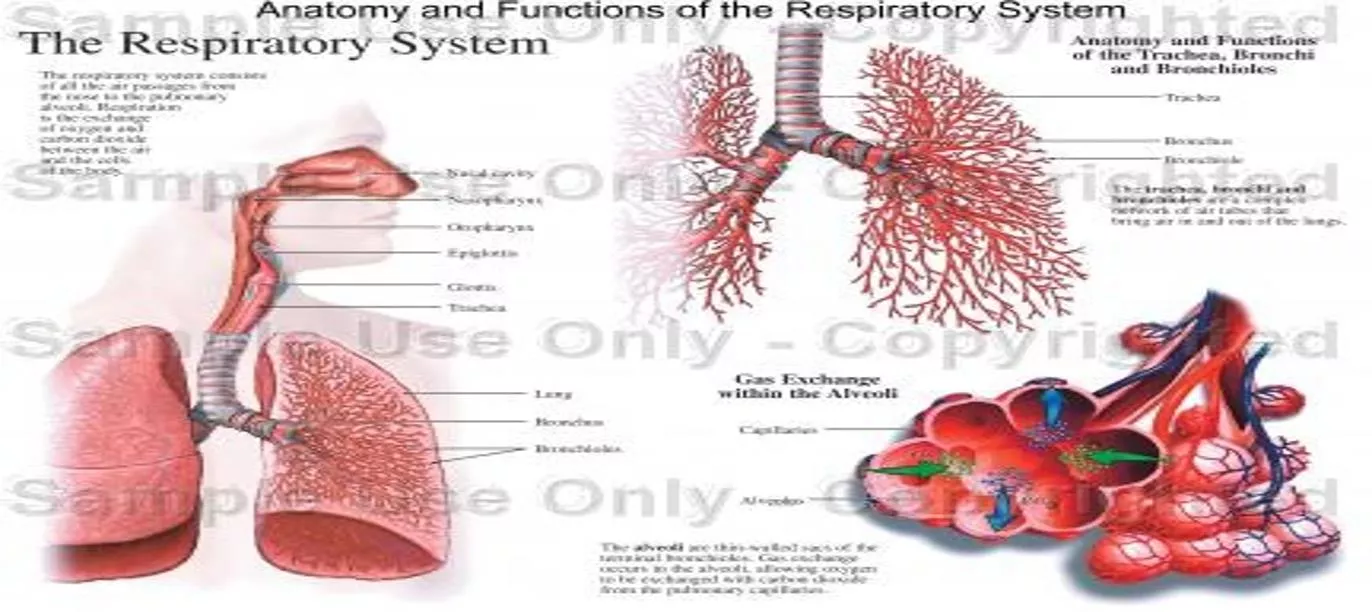
Functions
of the respiratory
system
include
•
Gas exchange
(respiratory
function).
•
Phonation:
is the
production of sounds
by the
movement of
air
through
the
vocal
cords
.
•
Pulmonary
defense
-
Immunoglobulin A
(IgA),
-
Alpha-1
antitrypsin
-
The
pulmonary
macrophages
in the
alveoli:
engulf
smaller particles which pass
through
the
muco-
cilliary barrier
filter
.
Dr.Aida
Korish (
akorish@ksu.edu.sa)
Cont..non respiratory
functions
of
lung
•
Angiotensin
I is
converted
to
angiotensin
II
with
the
help
of
angiotensin converting enzyme formed
by
the
lungs.
•
Regulating
the
acid-‐
base status
of
the
body
by
washing out
extra
carbon dioxide from
the
blood.
•
Secretion of important
substances like surfactant
.
Dr.Aida
Korish (
akorish@ksu.edu.sa)
Respiratory passages
(
airways)
Dr.Aida
Korish (
akorish@ksu.edu.sa)
Respiratory passages airways can
be
divided
into
Dr.Aida
Korish (
akorish@ksu.edu.sa)
receptors
for
smell
sensation.
Conducts
the
sound
during
sp
ee
ch.
Pr
o
tec
ti
ve
func
ti
o
n
by
c
o
ugh
and sneezing
reflexes.
II-‐
Respiratory
Zone
(Respiratory
unit)
•
Sta
r
ts fr
o
m n
o
se to the end
o
f
•
terminal
bronchioles.
•
Help
warming, humidification,
filtration
of
inspired
air.
Contains
the
olfactory
Includes:
Respiratory
bronchioles,
alveolar
ducts,
alveolar
sacs,
alveoli
•
Function
in gas
exchange.
I-‐
Conductive
Zone
Dr.Aida
Korish (
akorish@ksu.edu.sa)
Internal & External
Respiration
Dr.Aida
Korish (
akorish@ksu.edu.sa)
External
respiration
3
major functional
events
occurs
during
it:
1-‐Pulmonary
ventilation
: inward
and
outward
movement of
air
between
lung and
atmosphere.
2-‐
Diffusion
of oxygen
and CO2
between
the
alveoli
and
the
pulmonary
capillary
blood
3-‐
Transport
of
O2 &
Co2
in the
blood
and
body
fluids to
and
from
the
cells
Respiration could
be
either
Resting
:
normal breathing during resting
conditions.
Forced
(maximal):
during exercise,
in
patients
with
asthma,
allergy,…
Dr.Aida
Korish (
akorish@ksu.edu.sa)
Lining cells of the
alveoli
Dr.Aida
Korish (
akorish@ksu.edu.sa)
1-‐
Type I
alveolar
cells
( type I
pneumocytes)
2-‐
Type II
alveolar
cells
( type II
pneumocytes)
(Secrete
surfactant)
3-‐
Alveolar
macrophages
Surface
Tension
•
H
2
O
molecules at
the
surface
are attracted
to
other
H
2
O
molecules
by
attractive
forces that resist distension
called surface
tension.
•
Surface tension tends
to
oppose
alveoli
expansion.
•
Pulmonary surfactant
reduces surface
tension.
Dr.Aida
Korish (
akorish@ksu.edu.sa)
Surfactant
•
Surfactant is a
complex
substance
containing
phospholipids
and a number
of
apoproteins.
•
Secreted by the
Type
II
alveolar
cells. The earliest
detection from
fetal
alveoli
begins
between
6-‐7
th
month
but this
could
be
delayed
in
others
to
wk
35
of
intrauterine
life.
•
Surfactant reduces surface
tension throughout
the
lung,
prevents alveolar collapse,
decreases
airway
resistance and the
work of
breathing.
Dr.Aida
Korish (
akorish@ksu.edu.sa)
Cont…surfactant
•
Deficiency
in
premature babies cause respiratory
distress syndrome
of the
new
born (RDS) (
hyaline
membrane
disease)
•
Smoking in adult, hypoxia or hypoxemia
(low
oxygen
in the
arterial
blood) or both,
decrease
the
secretion
of
surfactant and cause adult respiratory
distress
syndrome.
Dr.Aida
Korish (
akorish@ksu.edu.sa)
Innervations
of
lungs
and
bronchi
•
Is by
autonomic
nerves.
•
Sympathetic
stimulation
causes
dilatation
of
the
bronchi
•
Parasympathetic stimulation
causes
constriction of
the
bronchi.
•
Locally
secreted
factors
:
histamine, slow reacting
substances
of anaphylaxis
(SRSA) by mast cells, due
to
allergy ( as in
patients with
asthma)
often
cause
bronchiolar
constriction
and
increase
airway
resistance.
Dr.Aida
Korish (
akorish@ksu.edu.sa)
Dr.Aida
Korish (
akorish@ksu.edu.sa)
Mechanics
of
pulmonary
ventilation
Learning
Objectives
•
By the
end
of this
lecture
you will be
able
to:
1
List the
muscles
of
respiration and describe their roles
during
inspiration and
expiration.
2
Understand
the
importance
of the following
pressures
in
respiration: atmospheric,
alveolar,
intrapleural,
and
transpulmonary.
3
Explain
why
intrapleural pressure
is
always subatmospheric
under normal conditions, and
the
significance
of the thin
layer
of the
intrapleural
fluid
surrounding
the lung.
4
Define lung compliance
and
list
the
determinants
of
compliance
.
Dr.Aida
Korish (
akorish@ksu.edu.sa)
Dr.Aida
Korish (
akorish@ksu.edu.sa)
Respiratory
muscles
Inspiratory
muscles
(resting-‐
forced)
Expiratory
muscles
(forced
expiration-‐
muscles that depress
the rib
cage)
Dr.Aida
Korish (
akorish@ksu.edu.sa)
Deep Forceful
Breathing
•
Deep
Inspiration
–
During
deep forceful inhalation accessory muscles
of
inspiration participate
to
increase size
of the
thoracic
cavity
•
Sternocleidomastoid
–
elevate
sternum
•
Scalene
–
elevate
first two
ribs
•
Pectoralis
minor –
elevate
3
rd
–5
th
ribs
•
Deep
Expiration
–
Expiration
during
forceful breathing
is
active
process.
–
Muscles
of
exhalation increase pressure
in
abdomen and
thorax
•
Abdominal
muscles.
•
Internal
intercostals.
Dr.Aida
Korish (
akorish@ksu.edu.sa)
Air will flow from a region of high pressure to one of low
pressure-- the bigger the
difference,
the faster the
flow
Dr.Aida
Korish (
akorish@ksu.edu.sa)
Pressure changes in the lungs during
breathing
1-Intra-alveolar (intrapulmonary
pressure
Between
breathes
=
zero
pressure
During
inspiration
=
(-1 mmHg).
air
(tidal volume)
flow from outside
to
inside the
lungs).
At the
end
of
inspiration
=
zero.
air
flow
stops.
During expiration
= (+1 mmHg).
air
flow out of the
Lungs
Dr.Aida
Korish (
akorish@ksu.edu.sa)
•
2-Intrapleural
pressure
(IPP):
Pressure
in the
pleural
space
is
negative
with
respect
to
atmospheric pressure at
the
end
of
normal expiration(
-5cmH2O).
•
Why
negative??:
1
The
lung's
elastic
tissue
causes
it to
recoil,
while
that
of the
chest wall causes
it to
expand. Because
of
these
2
opposing
forces
the
pressure
in the
pleural cavity
becomes
negative.
2
The pleural space
is a
potential space, empty
due to
continuous suction
of fluids by
lymphatic
vessels.
Dr.Aida
Korish (
akorish@ksu.edu.sa)
Values
of
IPP
•
(-5)
cm
H2O
during
resting
position
between
breathes, and
it
becomes
more –ve (-7.5)
cm
H2O
during resting
inspiration.
•
Forced
ventilation
Insp. :-20 to - 40
cm
H2O
Exp.
: + 30
cm
H2O
Dr.Aida
Korish (
akorish@ksu.edu.sa)
3-Transpulmonary
pressure
(TPp)
(Extending
Pressure)
•
The
difference
between
the
alveolar
pressure
(Palv)
and the
pleural
pressure(Ppl).
TPp
=
Palv-Ppl
•
It is a
measure
of the
elastic
forces
in the lungs that
tend
to
collapse
the lungs (
the
recoil
pressure).
It
prevents
lung
collapse.
•
The bigger
the volume of the lung the
higher
will be
its
tendency
to
recoil.
Dr.Aida
Korish (
akorish@ksu.edu.sa)
(Compliance
of
the lung
) in a single
respiratory
cycle
Dr.Aida
Korish (
akorish@ksu.edu.sa)
•
Is
defined as,
the
ratio
of the
change
in the lung
volume
produced per
unit
change
in the
distending
pressure.
•
The extent
to
which
the lungs
expand
for
each
unit
increase
in the
transpulmonary
pressure.
•
CL=
Volume
change
(∆
V)
Transpulmonary
pressure change
(∆
P)
•
CL
=
(∆
V)
(∆
P)
Dr.Aida
Korish (
akorish@ksu.edu.sa)
Cont…compliance of
lung
•
For both lungs in
adult
= 200 ml
of
air /cm
H20.
•
For lungs
and thorax together
=
110
ml/cm
H20.
•
Is
reduced
in
pulmonary
fibrosis ,
pulmonary edema, diseases
of the
chest wall
( kyphosis,
scoliosis)
•
Emphysema increases
the
compliance
of the lungs
because
it
destroys
the
alveolar septal
tissue
rich
with
elastic
fibers
that
normally
opposes
lung
expansion.
Dr.Aida
Korish (
akorish@ksu.edu.sa)
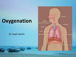
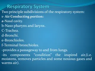
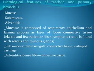
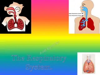
Within the complex respiratory system, the goal is to provide oxygen to tissues and remove CO2. It consists of airways, muscles, and centers. Functions include gas exchange, phonation, and pulmonary defense. The system also performs non-respiratory functions like converting Angiotensin I to II, regulating acid-base status, and secretion of surfactant. An in-depth look at the structures, functions, and organization of the system is provided. Key learning objectives encompass understanding the zones of airways, respiration types, and the system's broader functions beyond respiration.

Download Presentation

Please find below an Image/Link to download the presentation.
The content on the website is provided AS IS for your information and personal use only. It may not be sold, licensed, or shared on other websites without obtaining consent from the author. Download presentation by click this link. If you encounter any issues during the download, it is possible that the publisher has removed the file from their server.
E N D
Presentation Transcript
Respiratory Physiology Dr. Aida Korish Associate Prof.Physiology KSU Dr.Aida Korish ( akorish@ksu.edu.sa)
The main goal of respiration is to 1 Provide oxygen to tissues 2 Remove CO2 Respiratory system consists of: passages (airways) muscles centers Dr.Aida Korish ( akorish@ksu.edu.sa)
Functions and organization of the respiratory system Dr.Aida Korish ( akorish@ksu.edu.sa)
Learning Objectives By the end of this lecture you will be ableto:- 1-Describe the structures and functions of the conductive and respiratory zones of airways. 2- Understand the difference between internal and externalrespiration. 3- Understand the functions of the respiratory system, including non- respiratory functions, like clearance mechanism by mucus and cilia, production of surfactant and its physiologicalsignificance. Dr.Aida Korish ( akorish@ksu.edu.sa)
Functions of the respiratory system include Gas exchange (respiratory function). Phonation: is the production of sounds by the movement of air through the vocal cords. Pulmonary defense - Immunoglobulin A(IgA), - Alpha-1 antitrypsin - The pulmonary macrophagesin the alveoli: engulf smaller particles which pass through themuco- cilliary barrier filter. Dr.Aida Korish ( akorish@ksu.edu.sa)
Cont..non respiratory functions of lung Angiotensin I is converted to angiotensin II with the help ofangiotensin converting enzyme formed by the lungs. Regulating the acid- base status of the body by washing out extra carbon dioxide from the blood. Secretion of important substances like surfactant . Dr.Aida Korish ( akorish@ksu.edu.sa)
Respiratory passages ( airways) Dr.Aida Korish ( akorish@ksu.edu.sa)
Respiratory passages airways can be divided into Dr.Aida Korish ( akorish@ksu.edu.sa)
II- Respiratory Zone (Respiratory unit) I- Conductive Zone Starts from nose to the end of terminalbronchioles. Help warming, humidification, filtration of inspiredair. Contains the Includes: Respiratory bronchioles, alveolar ducts, alveolar sacs, alveoli Function in gasexchange. olfactory receptors for smellsensation. Conducts the sound during speech. P r otective function by cough and sneezingreflexes. Dr.Aida Korish ( akorish@ksu.edu.sa)
Internal & External Respiration Dr.Aida Korish ( akorish@ksu.edu.sa)
Externalrespiration 3 major functional events occurs during it: 1- Pulmonary ventilation: inward and outward movement of air between lung and atmosphere. 2- Diffusion of oxygen and CO2 between the alveoli and the pulmonary capillary blood 3- Transport of O2 & Co2 in the blood and body fluids to and from thecells Respiration could be either Resting: normal breathing during resting conditions. Forced (maximal): during exercise, in patients with asthma, allergy, Dr.Aida Korish ( akorish@ksu.edu.sa)
Lining cells of thealveoli 1- Type I alveolar cells ( type I pneumocytes) 2- Type II alveolar cells ( type II pneumocytes) (Secretesurfactant) 3- Alveolar macrophages Dr.Aida Korish ( akorish@ksu.edu.sa)
Surface Tension H2O molecules at the surface are attracted to other H2O molecules by attractive forces that resist distension called surface tension. Surface tension tends to oppose alveoli expansion. Pulmonary surfactant reduces surface tension. Dr.Aida Korish ( akorish@ksu.edu.sa)
Surfactant Surfactant is a complex substance containing phospholipids and a number of apoproteins. Secreted by the Type II alveolar cells. The earliest detection from fetal alveoli begins between 6- 7th month but this could be delayed in others towk 35 of intrauterine life. Surfactant reduces surface tension throughout the lung, prevents alveolar collapse, decreases airway resistance and the work ofbreathing. Dr.Aida Korish ( akorish@ksu.edu.sa)
Contsurfactant Deficiency in premature babies cause respiratory distress syndrome of the new born (RDS) ( hyaline membrane disease) Smoking in adult, hypoxia or hypoxemia (low oxygen in the arterial blood) or both, decrease the secretion of surfactant and cause adult respiratory distress syndrome. Dr.Aida Korish ( akorish@ksu.edu.sa)
Innervations of lungs and bronchi Is by autonomicnerves. Sympathetic stimulationcauses dilatationof the bronchi Parasympathetic stimulation causes constriction of the bronchi. Locally secreted factors :histamine, slow reacting substances of anaphylaxis (SRSA) by mast cells, due to allergy ( as in patients with asthma) often cause bronchiolar constriction and resistance. increase airway Dr.Aida Korish ( akorish@ksu.edu.sa)
Mechanics of pulmonary ventilation Dr.Aida Korish ( akorish@ksu.edu.sa)
Learning Objectives By the end of this lecture you will be able to: 1 List the muscles of respiration and describe their roles during inspiration and expiration. 2 Understand the importance of the following pressures in respiration: atmospheric, transpulmonary. 3 Explain why intrapleural pressure is always subatmospheric under normal conditions, and the significance of the thin layer of the intrapleural fluid surrounding the lung. 4 Define lung compliance and list the determinants of compliance. alveolar, intrapleural, and Dr.Aida Korish ( akorish@ksu.edu.sa)
Respiratorymuscles Inspiratorymuscles (resting- forced) Expiratory muscles (forced expiration- muscles that depress the ribcage) Dr.Aida Korish ( akorish@ksu.edu.sa)
Deep Forceful Breathing Deep Inspiration During deep forceful inhalation accessory muscles of inspiration participate to increase size of the thoracic cavity Sternocleidomastoid elevate sternum Scalene elevate first two ribs Pectoralis minor elevate 3rd 5thribs Deep Expiration Expiration during forceful breathing is active process. Muscles of exhalation increase pressure in abdomen and thorax Abdominal muscles. Internal intercostals. Dr.Aida Korish ( akorish@ksu.edu.sa)
Air will flow from a region of high pressure to one of low pressure-- the bigger the difference, the faster the flow Dr.Aida Korish ( akorish@ksu.edu.sa)
Pressure changes in the lungs during breathing 1-Intra-alveolar (intrapulmonary pressure Between breathes =zero pressure During inspiration = (-1 mmHg). air (tidal volume) flow from outside to inside the lungs). At the end of inspiration =zero. air flow stops. During expiration = (+1 mmHg). air flow out of the Lungs Dr.Aida Korish ( akorish@ksu.edu.sa)
2-Intrapleural pressure (IPP): Pressure in the pleural space is negative with respect to atmospheric pressure at the end of normal expiration( -5cmH2O). Why negative??: 1The lung's elastic tissue causes it to recoil, while that of the chest wall causes it to expand. Because of these 2 opposing forces the pressure in the pleural cavity becomes negative. 2The pleural space is a potential space, empty due to continuous suction of fluids by lymphatic vessels. Dr.Aida Korish ( akorish@ksu.edu.sa)
Values ofIPP (-5) cm H2O during resting position between breathes, and it becomes more ve (-7.5) cm H2O during resting inspiration. Forced ventilation Insp. :-20 to - 40 cm H2O Exp. : + 30 cm H2O Dr.Aida Korish ( akorish@ksu.edu.sa)
3-Transpulmonary pressure (TPp) (Extending Pressure) The difference between the alveolar pressure (Palv) and the pleural pressure(Ppl). TPp = Palv-Ppl It is a measure of the elastic forces in the lungs that tend to collapse the lungs (the recoil pressure). It preventslung collapse. The bigger the volume of the lung the higher will be its tendency to recoil. Dr.Aida Korish ( akorish@ksu.edu.sa)
(Compliance ofthe lung) in a single respiratory cycle Dr.Aida Korish ( akorish@ksu.edu.sa)
Is defined as, the ratio of the change in the lung volume produced per unit change in the distending pressure. The extent to which the lungs expand for each unit increase in the transpulmonary pressure. CL= Volume change ( V) Transpulmonary pressure change ( P) CL = ( V) ( P) Dr.Aida Korish ( akorish@ksu.edu.sa)
Contcompliance of lung For both lungs in adult = 200 ml of air /cm H20. For lungs and thorax together = 110 ml/cm H20. Is reduced in pulmonary fibrosis , pulmonary edema, diseases of the chest wall ( kyphosis, scoliosis) Emphysema increases the compliance of the lungs because it destroys the alveolar septal tissue rich with elasticfibers that normally opposes lung expansion. Dr.Aida Korish ( akorish@ksu.edu.sa)


")


")


:")


 in a single respiratory cycle")