Understanding the Respiratory System Components and Functions
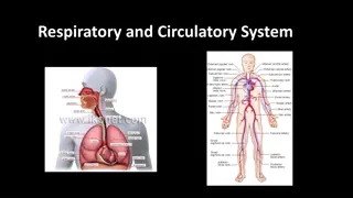
The respiratory system consists of two primary subdivisions: the Air Conducting portion and the Respiratory portion. The Air Conducting portion includes nasal cavity, naso-pharynx, larynx, trachea, bronchi, bronchioles, and terminal bronchioles, providing a pathway to and from the lungs while conditioning the inspired air. The Respiratory portion comprises respiratory bronchioles, alveolar ducts, and alveolar sacs for gas exchange. Structural components like cartilage, elastic fibers, and smooth muscles help keep the air passages open. The system functions to supply oxygen for cellular oxidation and remove carbon dioxide. Various cells like ciliated cells, goblet cells, and basal cells contribute to maintaining the respiratory epithelium.

Uploaded on Sep 23, 2024 | 0 Views
Download Presentation

Please find below an Image/Link to download the presentation.
The content on the website is provided AS IS for your information and personal use only. It may not be sold, licensed, or shared on other websites without obtaining consent from the author. Download presentation by click this link. If you encounter any issues during the download, it is possible that the publisher has removed the file from their server.
E N D
Presentation Transcript
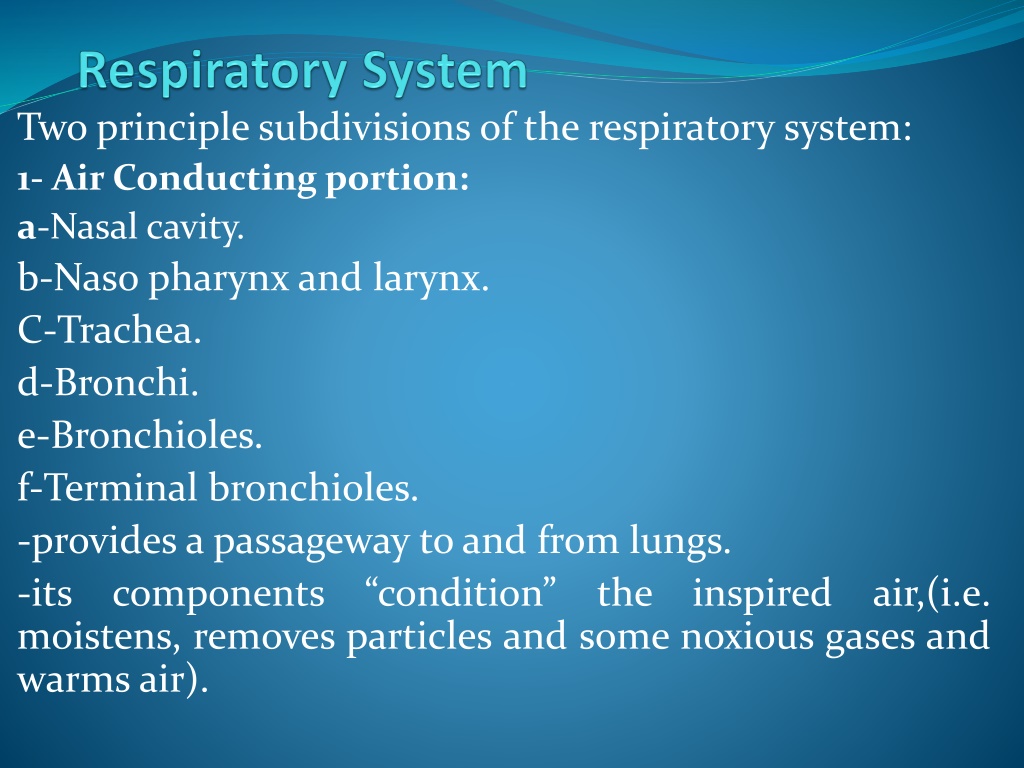
Two principle subdivisions of the respiratory system: 1- Air Conducting portion: a-Nasal cavity. b-Naso pharynx and larynx. C-Trachea. d-Bronchi. e-Bronchioles. f-Terminal bronchioles. -provides a passageway to and from lungs. -its components condition moistens, removes particles and some noxious gases and warms air). the inspired air,(i.e.
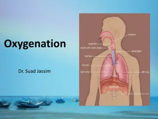
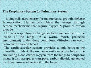
2-Respiratory portion: a-Respiratory bronchioles. b-Alveolar ducts. c-Alveolar sacs and alveoli for gas exchange. _Function of respiratory system: The function of the respiratory system is to provide molecular oxygen for cellular oxidation and to remove carbon dioxide generated as a waste product of cell metabolism. There must be vascular or circulatory transport and air/gas transport.
_The structural components that keep the air passage wayalwaysopened: There are three structures in the wall of respiratory tract, they keep the airway patent and opened; they are: 1-Cartilage (Hyalinecartilage) : support the wall, prevent collapse of the lumen and give rigidity to the wall. 2-Elastic fiber: Provide flexibility to wall; also permit elongation of the conducting portion. 3-Smooth muscle: Reduce the diameter of the tube and regulates the air flow.
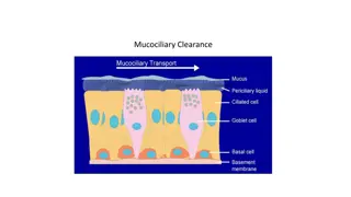
_The air must be condition before it reaches the respiratory portion, so the wall of conducting portion; have many structural features to serve these functions: -The mucosa is epithelium and lamina propria, most parts of conducting portion are lined by (epithelium called respiratory epithelium stratified ciliated columnar epithelium with goblet cells. -The cilia =can push the dust particles and mucus by its motion toward the pharynx either to be swilled or thrown out. -Goblet cells and mucous glands=cover the epithelium with protective mucous secretion that trap the dust particles, and moisten the air. which is pseudo
-The lamina propria contains serous (keep the epithelium moist, and humidify the air) and mucous glands, and numerous blood vessels (warm theair). _Cellsof respiratoryepithelium: It is composed of five types of cell: 1-Ciliated cells: Are tall columnar cells have cilia, basal located nucleus. 2-Goblet cells. 3-Brush cells (intermediatecells): Have microvilli, transform in to either ciliated cells or goblet cells. 4-Basal cells: Are short cells lie on the basement membrane but don t reach the surface, they are stem cells from which other cell types are developed.
5-Granular cells (neuro endocrine cells): are rounded cells have dark stained nucleus, and secretary granules. _Nasal Cavity: Is divided in to right &left, by nasal septum and composed of three parts: Vestibule region: is widest part continuous with external skin, has stiff thick hairs to remove the dust particles from inspired air, and sweat glands. The lamina propria contains serous mucus glands, and numerous blood vessels. Respiratory region: is lined by pseudo stratified ciliated columnar with goblet cells. Lamina propria contains serous; mucous glands, and blood vessels.
Olfactory region: is located in the roof of nasal cavity; lined by epithelium thicker than that of respiratory area, it is pseudo stratified columnar epithelium, composed of 3 typesof cells: *Supporting cells. *Basal cells. *Olfactorycells. _Supporting cells: are tall cells, columnar in shape narrow at base in contact with basement membrane, wide near the lumen that have microvilli, oval nucleus, cytoplasm has secretory granules, these cells give physical support or they may be phagocytic cell. _Basal cells: short, conical shape cells rest on the basement membrane butdon t reach to the surface, the
Cell has cytoplasmic processes, and has oval nucleus. They are stem cells from which new olfactory cells or supporting are developed. _Olfactorycells(sensorycells): -spindle- shape cells, rounded nucleuscentrally located. -have two process, the (Axon) toward the lamina propria, and (Dendrite) toward the surface, that project above the epithelium as (knob-like) structure called (olfactory vesicle) from which about (10-25) non-motile small process called (olfactory hairs) actas receptive structure. -Brown gland is tubulo-acinar gland (serous gland), found in the lamina propria that contains collagen fiber, blood and lymphaticcapillaries. The secretion is carried to
Surface by duct in the olfactory epithelium that act as solvent to substances. -para nasal sinuses-they sphenoid, frontal sinuses. Communicated with nasal cavity so they are lined by respiratory epithelium, they increase the surface area for warming and moistening the airalso play role in sound. _Nasopharynx: The epithelium is respiratory epithelium, lamina propria contains serous and mucous gland. _Larynx: Is rigid tube about 4 cm in length, its wall contains, cartilage, muscle and mucous membrane. are maxillary, ethmoid,
-the cartilage of hyaline type is thyroid, cricoids and arytenoids cartilage. -the elastic cartilage epiglottis, is valve-like structure covered by mucous membrane on both sides. -anterior surface stratified squamous epithelium non- keratinized. -posterior surface pseudostratified ciliated columnar epithelium with goblet cells. Vocal cords: - are two paired of folds extend in to the lumen of larynx from the mucosa below the epiglottis. -the upper fold (false vocal cords) lined by respiratory epithelium.
-the lower fold(true vocal cord) lined by stratified sqaumous epithelium. -vocal ligament and bundle of skeletal muscle (vocal muscle). _Trachea: -is rigid tube about (10-12) cm long (2-3) cm in diameter. -has about (16-20) rings of (c-shaped) hyaline cartilage. -the perichondrium of each rings is connected to other ring by fibro-elastic connective tissue. -the free ends of cartilage are joined by smooth muscle (trachealis muscle).


:")


 lined by stratified")