Understanding Connective Tissue Wrappings and Skeletal Muscle Structure
Exploring the connective tissue wrappings and attachments of skeletal muscles, this content highlights the cord-like structures, collagen fibers, and their role in connecting muscle bellies to bones. It also delves into the surrounding connective tissues such as fascia, epimysium, perimysium, and endomysium, providing insights into their functions in supporting muscle cells. Additionally, the microscopic anatomy of skeletal muscles is discussed, focusing on sarcolemma and myofibrils. The content concludes with a creative Play Doh Model Challenge to replicate skeletal muscle structure using different colors of modeling clay.

Download Presentation

Please find below an Image/Link to download the presentation.
The content on the website is provided AS IS for your information and personal use only. It may not be sold, licensed, or shared on other websites without obtaining consent from the author. Download presentation by click this link. If you encounter any issues during the download, it is possible that the publisher has removed the file from their server.
E N D
Presentation Transcript
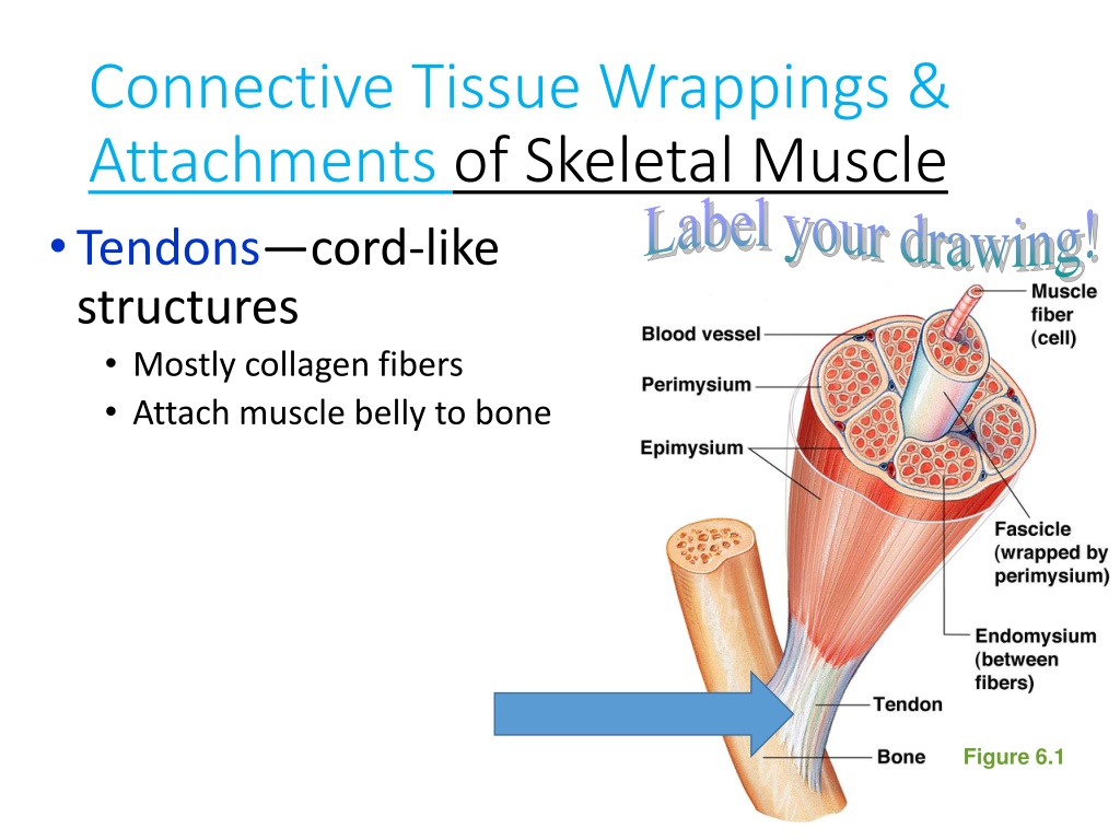
Connective Tissue Wrappings & Attachments of Skeletal Muscle Tendons cord-like structures Mostly collagen fibers Attach muscle belly to bone Label your drawing! Figure 6.1
Connective Tissue Wrappings & Attachments of Skeletal Muscle Cells are surrounded & bundled by connective tissue Fascia tough connective tissue, outside of the epimysium of the muscle belly, that creates a 3-D web around every muscle Figure 6.1
Connective Tissue Wrappings & Attachments of Skeletal Muscle Cells are surrounded & bundled by connective tissue Epimysium covers the entire skeletal muscle (muscle belly) Label your drawing & Figure 6.1
Connective Tissue Wrappings & Attachments of Skeletal Muscle Cells are surrounded & bundled by connective tissue Perimysium wraps around a fascicle (bundle) of muscle fibers Label your drawing & Figure 6.1
Connective Tissue Wrappings & Attachments of Skeletal Muscle Cells are surrounded & bundled by connective tissue Endomysium encloses a single muscle fiber Label your drawing & Figure 6.1
Microscopic Anatomy of Skeletal Muscle Label your drawing & use your last color to identify the myofibrils! Sarcolemma specialized plasma membrane that surrounds a myofibril Myofibrils long organelles inside muscle cell
Per. 1 - The end REVIEW of SKELETAL MUSCLE STRUCTURE: REVIEW of SKELETAL MUSCLE STRUCTURE: Play Doh Model Challenge Using the 4 colors of modeling clay, develop a mini replica of skeletal muscle, including 1 muscle belly At least 3 fascicles At least 3 muscle fibers within each fascicle At least 3 myofibrils within each muscle fiber Muscle belly Fascicles Muscle fibers Myofibrils
Per. 1 - The end REVIEW of SKELETAL MUSCLE STRUCTURE: REVIEW of SKELETAL MUSCLE STRUCTURE: Play Doh Model Challenge Follow Up Questions: Which colors did you use to represent each structure? Which structure did you construct first? Explain why you did this Which part of the muscle wrapping was the most complicated to build? Explain your answer Draw and color your muscle structure and include labeling of the wrappings and their functions Muscle belly Fascicles Muscle fibers Myofibrils
Today in Human Anatomy... Pick up: Skeletal Muscle Crossword Textbook Have out: Mr. Muscle Man Muscle Structure diagram Week #6 (11/16-11/20) Warm Up Fri, 11/20: - Review of Muscle #2 & Muscle organization Anatomy Fun Fact: As the body gets cold, heat is generated due to shivering which causes rapid muscle contractions. Homework: 1. Muscles Quiz #2 (Identification) Mon, 11/23 2. Muscular Sys. Rev. packet Wed, 11/25 3. Muscle System Quiz #1 (Structure, Function & Organization) Mon, 11/30 Agenda: 1. Muscular System Latin Quiz 2. Skeletal Muscle Crossword Puzzle due Monday 11/23 Learning Goal: I will be able to describe the structure & function of the Muscular System.
Skeletal Muscle Organization Name the following structures & connective tissue wrappings of skeletal muscle. B C (structure) A (wrapping) G (wrapping) E (wrapping) D (structure) F (structure) I (wrapping of H) H (structure)
Per. 3 needs; go over WU & collect, The end Bionics Photo Gallery National Geographic: Bionics QUIETLY read the article Bionics . Annotate the article as you please (symbols, writing in margins, highlighting). Prompt: We are giving people tools. They are better than what previously existed. But they are still crude, like a hammer, compared with the complexity of the human body. They can t hold a candle to Mother Nature. Using your knowledge of the interdependence of organ systems in the human body & how skeletal muscle is organized & using direct information about bionics in the article, discuss why this quote is so truthful. 3-paragraph response (opening, body, closing) STAPLE your annotated article to your written response & turn in to the Hmwk Bin! DUE by the END of the PERIOD TODAY!