Understanding the Histological Structure of Muscle Cells

This presentation delves into the histological structure of three types of muscle cells - skeletal, cardiac, and smooth muscles. Learn about their differences, such as striation, voluntary vs. involuntary control, and cellular organization. Discover the components of muscular tissue, the coverings of skeletal muscle, and the microscopic features of skeletal muscle fibers. Unravel the intricacies of myofibrils within skeletal muscles and their role in muscle contraction.

Download Presentation

Please find below an Image/Link to download the presentation.
The content on the website is provided AS IS for your information and personal use only. It may not be sold, licensed, or shared on other websites without obtaining consent from the author. Download presentation by click this link. If you encounter any issues during the download, it is possible that the publisher has removed the file from their server.
E N D
Presentation Transcript
Integrated Muscle Red: important. Black: in male|female slides. Gray: notes|extra. Editing File Editing File
OBJECTIVES Identify and describe the histological structure of the three types of muscle cells and list the differences between them. Histology team 437 | MSK block | Lecture two
Extra picture for explanation only Histology team 437 | MSK block | Lecture two
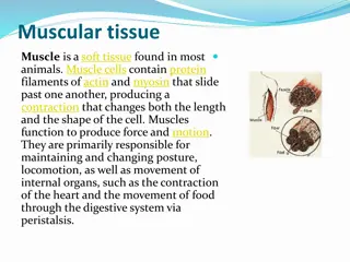
MUSCULAR TISSUE =carco=myo=mysium Made of elongated muscle cells (fibers) Skeletal types of muscles striated, voluntary The Cells are the fibers (muscle fibers) Cardiac striated, involuntary The fibers are segmented to cells Smooth non-striated, involuntary The Cells are the fibers Muscle cell is only cell that we can see by naked eye Muscle supply by somatic nerve Histology team 437 | MSK block | Lecture two
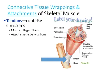
SKELETAL MUSCLE The whole muscle is covered by a connective tissue covering called the epimysium consist of dense collagen irregular connective tissue Consists of parallel skeletal muscle fibers, arranged in bundles, separated by connective tissue septa called the perimysium consist of loose connective tissue The individual fibers are separated by connective tissue called endomysium. Mysium = Flesh Epi = Over Peri = Surrounding Histology team 437 | MSK block | Lecture two
Skeletal Muscle Fibers under Microscope L.M. Picture E.M. Picture Cylindrical in shape. Non-branched. Covered by a clear cell membrane, the sarcolemma. Multinucleated: nuclei are multiple and are peripherally located (close to the sarcolemma). Cytoplasm (sarcoplasm) is acidophilic and shows clear transverse striations. Sarcoplasm contains: Parallel myofibrils. Numerous mitochondria, arranged in rows between the myofibrils. Well developed smooth endoplasmic reticulum (sarcoplasmic reticulum- SR). Myoglobin pigment. myoglobin is protein high affinity for oxygen Glycogen Glycogen is food for muscles Glycogen + Ca+ = contraction High concentration of Ca+ that release from smooth ER increase cause muscle contraction Histology team 437 | MSK block | Lecture two
Myofibrils of Skeletal Muscles E.M. Picture of Myofibrils: Contractile threads (organelles), arranged longitudinally in the sarcoplasm. Each myofibril shows alternating dark (A) and light bands (I). The (A) band shows a pale area in the middle (H band) which is divided by a dark line (M line). The (I) band shows a dark line in the middle (Z line). The sarcomere is the segment between 2 successive Z lines. It is the contractile unit of a myofibril. The myofibrils are formed of myofilaments (thick myosin and thin actin). The (A) band is formed of myosin myofilaments mainly and the terminal ends of actin myofilaments. The (I) band is formed of actin myofilaments. Sarcomere Z Z I I A The (A) band is dark because it contains both myosin and actin. The (I) band and the (H) zone are light because they have only one type of myofilaments (actin in I and myosin in H). Myosin Actin Histology team 437 | MSK block | Lecture two
CARDIAC MUSCLE Found in the myocardium. Striated and involuntary. Have only Endomysium. L.M. Picture E.M. Picture Cylindrical in shape. Intermediate in diameter between skeletal and smooth muscle fibers. Branch and anastomose. Covered by a thin sarcolemma. Mononucleated. Nuclei are oval and central. Sarcoplasm is acidophilic and shows non-clear striations (fewer myofibrils). Divided into short segments (cells) by the intercalated discs. Few myofibrils. Numerous mitochondria. Less abundant SR. Glycogen & myoglobin. Intercalated discs: are formed of the two cell membranes of 2 successive cardiac muscle cells, connected together by junctional complexes (desmosomes and gap junctions*). *Gap junctions allow communication and passage of impulses between cardiac muscle cells Histology team 437 | MSK block | Lecture two
SMOOTH MUSCLE Present in walls of blood vessels and viscera (digestive, urinary, genital .... etc). Non-striated and involuntary. Smooth muscle have dense body that help in contraction Gap junction found cardiac & smooth muscle L.M. Picture Fusiform in shape (spindle- shaped). Small diameter. Non-branched. Thin sarcolemma. Mononucleated. Nuclei are oval & central in position. Sarcoplasm is non-striated and acidophilic. E.M. Picture Sarcoplasm contains mitochondria and sarcoplasmic reticulum. Myosin & actin filaments are irregularly arranged (that s why no striations could be observed). Cells are connected together by gap junctions for cell communication. Histology team 437 | MSK block | Lecture two
REGENERATION OF MUSCLE (1) Skeletal muscle cells: - Can not divide. - Limited regeneration by satellite cells (stem cells on the muscle cell s surface) (2) Cardiac muscle cells: -No regenerative capacity. (3) Smooth muscle cells: - Can divide. - Regenerate from pericytes. active regenerative response. - mitosis for other healthy cell | stem cell Comparison between different types of muscle fibers SKELETAL Muscle attached to skeleton CARDIAC SMOOTH Myocardium of the heart Viscera, e.g. stomach Site Cylindrical Cylindrical Fusiform Shape Diameter Branching Striations Intercalated discs Nuclei Action Regeneration Largest Medium-sized Smallest Non-branched Branched Non-branched Clear Not clear Absent Absent Present Absent Numerous and peripheral One central nucleus One central nucleus Voluntary Involuntary Involuntary Histology team 437 | MSK block | Lecture two Limited No Active
QUESTIONS: Q1:What is the name of the dark line in the middle of The ( I ) band? A) M line B) Z line C) H line D) E line Q2:Which one of the following not striated? A) Cardiac muscle B) Skeletal muscle C) Smooth muscle D) All of them 5-A 4-D Q3: Intercalated discs is present in which of the following type of muscle fibers? A) Cardiac muscle B) Skeletal muscle C) Smooth muscle D) All of them 3-A 2-C 1-B Q4: one of the following is a common feature in both smooth and cardiac muscles? A) Multinucleated B) Steriation C) Fusiform cells D) Gap junctions Q5: Which one is never regenerate? A) Cardiac muscle B) Skeletal muscle C) Smooth muscle D)all of them Histology team 437 | MSK block | Lecture two
Q6: Whose have largest dimeter? A) Cardiac muscle B) Skeletal muscle C) Smooth muscle D) A&B Q7: Which of the following C.T. separates each individual skeletal muscle fibers? A) Sarcoplasm B) Perimysium C) Endomysium D) Epimysium 11-C Q8: the contractile unit of a myofibril? A) Sarcomere B) Sarcoplasm C) Sarcoplasmic reticulum D) Sarcolemma 10-B 9-D 8-A Q9: The shape of smooth muscle is? A) Cylindrical B) Circular C) Triangular D) Fusiform 7-C 6-B Q10: Sarcomere is the distance between two? A) M lines B) Z lines C) H lines D) E lines Q11:Which one of the following muscle fibers can divide? A) Cardiac muscle B) Skeletal muscle C) Smooth muscle D) All of them Histology team 437 | MSK block | Lecture two
Team members : Hussain Alkharboush Ebtesam Almutairi Shahad Alzahrani Tareq Allhaidan Marwah Alkhalil Rinad Alghoraiby Team leaders : Khalid Fayez Alshehri Rawan Mohammad Alharbi Twitter.com/Histology437 HistologyTeam437@gmail.com Histology team 437 | MSK block | Lecture two