Understanding the Muscular System and Its Anatomy
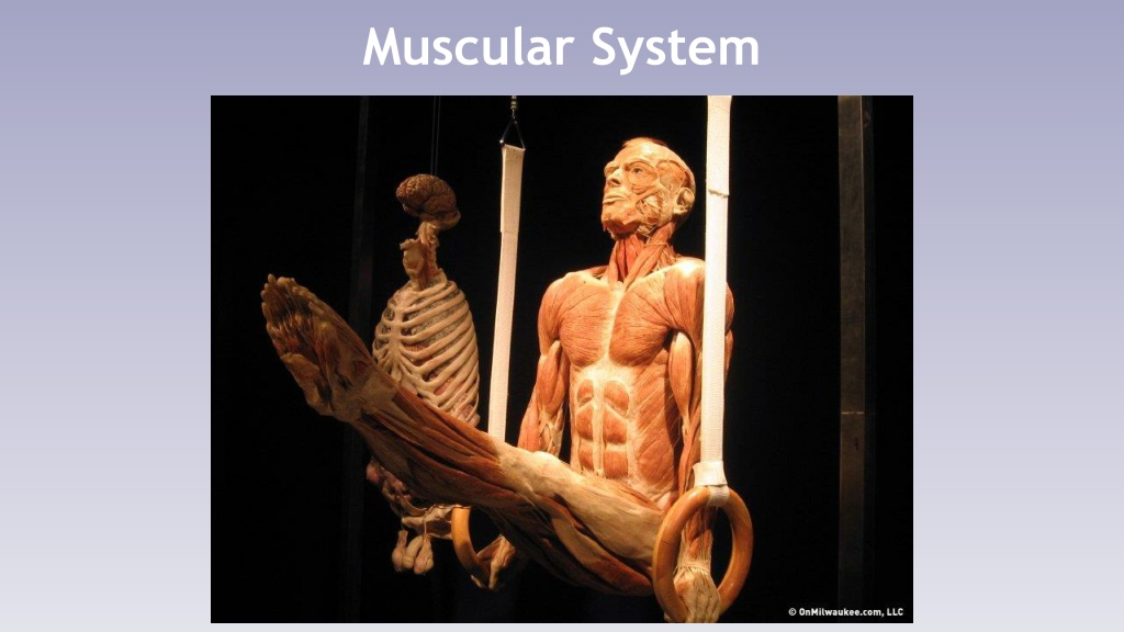
The muscular system is vital for movement, posture maintenance, and organ protection. It consists of skeletal muscles that contract to create movement, circular muscles called sphincters for controlling openings, and muscle fibers arranged in fascicles with collagen layers like epimysium and endomysium. Tendons and aponeurosis connect muscles to bones or other muscles. Muscle fibers are long cells, with the sartorius muscle containing some of the longest fibers in the body.

Download Presentation

Please find below an Image/Link to download the presentation.
The content on the website is provided AS IS for your information and personal use only. It may not be sold, licensed, or shared on other websites without obtaining consent from the author. Download presentation by click this link. If you encounter any issues during the download, it is possible that the publisher has removed the file from their server.
E N D
Presentation Transcript
Muscular System Functions Skeletal muscle pulls on the bones of the skeleton, creating movement. Even when not moving, skeletal muscle is partially contracted, maintaining tone and posture.
A wall of skeletal muscle provides protection for the organs of the abdominal cavity.
Circular muscles called sphincters control openings in the digestive and urinary systems. Muscle contractions generate heat, helping to maintain body temperature.
Anatomy of a Muscle Each muscle is covered with epimysium, a layer of collagen fibers that separates it from surrounding organs.
The muscle is made of bundles called fascicles. Each fascicle is divided by another layer called the perimysium. Nerves and blood vessels are also found here.
Individual muscle cells within a fascicle are called muscle fibers. The endomysium surrounds and each individual muscle fiber.
At each end of the muscle, the collagen fibers from all three layers come together to form one of two possible structures: A bundle of collagen fibers called a tendon, which attaches the muscle to a bone. A sheet of collagen fibers called a aponeurosis, which attaches the muscle to another muscle.
A ruptured tendon will detach a muscle completely from one of its bones, rendering it unusable.
Epimysium Perimysium Endomysium Tendon Fiber Muscle Fascicle
Muscle fibers are some of the longest cells in the body. Longest is in the sartorius muscle 60cm! Each fiber is multinucleate, meaning there are multiple nuclei in each cell, and amitotic, meaning they cannot divide.
The sarcolemma, the cell membrane of the muscle fiber, is covered with openings to a network of small tubules called T tubules. The sarcoplasmic reticulum is a special type of smooth ER that stores calcium ions that signal muscle contraction.
Each muscle fiber contains myofibrils, long filaments that have the ability to contract. Thin, light filaments, made of the protein actin. Thick, dark filaments, made of the protein myosin.
T-Tubules Sarcoplasmic Reticulum Sarcolemma Mitochondria Myosin Actin
Muscle Contraction Muscle contractions occur in the following sequence of events: A signal is passed through motor neuron to a muscle. This signal is sent to every fiber in the muscle simultaneously through the t-tubules. The sarcoplasmic reticulum releases calcium ions (Ca2+), initiating muscle contraction.
The calcium influx stimulates the myosin filaments to form connections to the actin filaments. The myosin filaments pull the actin filaments inward, causing the muscle to contract.
When a muscle contracts, it pulls bones closer together, creating movement. Contracted muscles become more visible because all of the volume (cytoplasm) is forced outward, creating a muscle belly.
The amount and force of muscle tension depends on: Frequency of simulation (form the central nervous system). Number of skeletal muscle fibers involved. The size of the muscle fibers larger fibers contain more myofibrils.
Muscle Stimulation Summation is the process of recruiting more muscle fibers to generate a greater force. Summation begins when a single electrical impulse from a neuron stimulates a twitch, a single stimulus- contraction-relaxation sequence in a muscle.
A twitch has three parts: The latent period, where the stimulus spreads through the muscle. The contraction phase, where action and myosin generate tension. The relaxation phase, where action and myosin uncouple and the muscle relaxes. Resting Phase Latent Period Contraction Phase
Continuous stimulation results in twitches overlapping, eventually producing a prolonged contraction called tetanus.
Energy for Muscle Contraction ATP is the direct unit of energy used by muscle fibers. ATP is converted to ADP when it is used. If the supply of ATP is exhausted, the muscle becomes fatigued and will not contract.
ATP is an unstable molecule, so cells only have small amounts available at any given moment. About 3 seconds worth. Creatine phosphate can be broken down to release high-energy phosphates, quickly recharging ATP. 8-1o seconds worth of contraction.
To continue contracting, muscle fibers must start using glucose. Glycolysis breaks down glucose, releasing two molecules of pyruvate and two molecules of ATP. Anaerobic, meaning no oxygen is used. Takes place in the cytoplasm. The next step depends on the oxygen levels in the muscle fiber.
If oxygen levels are high, cell respiration continues breaking down glucose into 34 molecules of ATP for every 1 molecule of glucose. Aerobic, meaning oxygen is required. Takes place in the mitochondria.
When oxygen levels are low, cells use fermentation reactions to recycle the unused products of glycolysis. Lactic acid is a byproduct of fermentation and can cause fatigue and soreness. It is gradually re-absorbed when oxygen levels return to normal.
When glucose levels run low, a polysaccharide called glycogen can be broken down to generate more. Glycogen is stored in the liver and muscles.
Muscle fatigue has two possible causes: Running out of glycogen. Limits available glucose to muscle cells. Hitting the wall. Insufficient oxygen levels. Forces muscle fibers to rely more on glycolysis and fermentation. Out of breath.
Types of Muscle Fibers Fast-twitch fibers are able to reach peak tension within 0.01 seconds or less of neural stimulation. Large in diameter. Densely packed with myofibrils (actin and myosin). Large glycogen reserves. Fewer mitochondria. Fast-twitch fibers produce the most tension, but get fatigued quickly.
Slow-twitch fibers can take three times as long to reach peak tension. Half the diameter of fast-twitch fibers. Increased network of capillaries, allowing for a greater and more reliable oxygen supply. Contain a special protein called myoglobin that reserves additional oxygen within the muscle. Higher numbers of mitochondria. Produce less power, but much more endurance.
Physical Conditioning Physical conditioning can focus on improving muscle force or endurance. Aerobic exercise focuses on improving endurance by improving oxygen intake and increasing glycogen storage. Jogging, distance swimming, etc. Anaerobic exercise improves strength by increasing the size of each muscle fiber. Each muscle fiber has more myofibrils.
Anabolic Steroids Anabolic steroids are chemical compounds that mimic the effects of testosterone. This increases protein synthesis in muscle fibers. As a hormone, testosterone affects many other tissues besides muscles, causing side effects: Increase in blood cholesterol. Acne High blood pressure Testicular atrophy Increase in male characteristics in women.
Muscular System Disorders Polio is a viral infection that can infects and destroys motor neurons, causing paralysis. Considered eradicated due to a vaccine.
ALS (Lou Gehrigs Disease) Amyotrophic Lateral Sclerosis (ALS), also known as Lou Gehrig s Disease, is a genetic neurodegenerative disease that damages motor neurons of the peripheral nervous system. Causes muscle atrophy due to disuse.
Muscular Dystrophy Muscular dystrophy is a group of genetic degenerative disorders that directly affects muscle tissue, causing it to atrophy.
How Are Muscles Named? Some muscles are named based on the direction of their fibers. Rectus means straight. Rectus abdominis. Oblique means diagonally arranged. External abdominal oblique.
Muscles within a group may have different names based on their size. Maximus and longus indicates a larger muscle. Minimus and brevis indicate a smaller muscle.
Muscles may also be named based on their relative location to other muscles. Medial means towards the midline of the body. Lateral means towards the sides of the body.
Prefixes like bi- and tri- may be used to indicate multiple heads or attachment sites. Bi Two attachment sites. Tri Three attachment sites.
Muscles may also be named based on their origin and insertion bones. The origin is an attachment to a immoveable bone. The insertion is an attachment to an movable bone.
If a muscle resembles a shape, it can be named after that shape. Delta is a Greek letter shaped like a triangle. Trapezius is shaped like a trapezoid. Serratus means saw- toothed.
Finally, muscle names may indicate a specific action they perform. Flex means to bend a joint. Extend means to straighten a joint.
Zygomatic Bone Temporalis Frontalis Orbicularis Oculi Zygomaticus Trapezius Orbicularis Oris Sternocleidomastoid Buccinator Masseter
Head and Neck Muscles The frontalis raises the eyebrows. The masseter and temporalis both elevate the mandible. Chewing muscles The buccinator flattens the cheeks during chewing, holding them against the teeth. The orbicularis oculi performs all eyelid movements, including opening, closing, blinking, etc. The orbicularis oris closes the mouth with the lips. The zygomaticus raises the corners of the mouth when smiling. The sternocleidomastoid rotates the head and flexes the neck.
Sternocleidomastoid Trapezius Clavicle Sternum Pectoralis Major Deltoid Serratus Anterior Latissimus Dorsi Internal Abdominal Oblique External Abdominal Oblique Rectus Abdominis
Muscles of the Trunk The pectoralis major adducts the humerus. The rectus abdominis flexes the vertebral column and compresses the contents of the abdomen. The pushing muscle of defecation, childbirth, and forced breathing. The transversus abdominis also performs this action. The external and internal obliques rotate the trunk.
Sternocleidomastoid Trapezius Deltoid Teres Major Infraspinatus Latissimus Dorsi External Oblique
Muscles of the Dorsal Trunk The trapezius elevates and depresses the scapula. The latissimus dorsi adducts the humerus. The deltoid abducts the arm.


")