Understanding Electromyography (EMG) and Motor Neurons


What is an EMG?


What is an EMG?
●
EMG stands for
electromyography
, which is an electrodiagnostic
technique for evaluating and recording the electrical activity produced
by skeletal muscles.
○
We use them in order to assess the health of muscles and the nerve cells that help
control them.
https://www.hopkinsmedicine.org/health/treatment-tests-and-
therapies/electromyography-emg
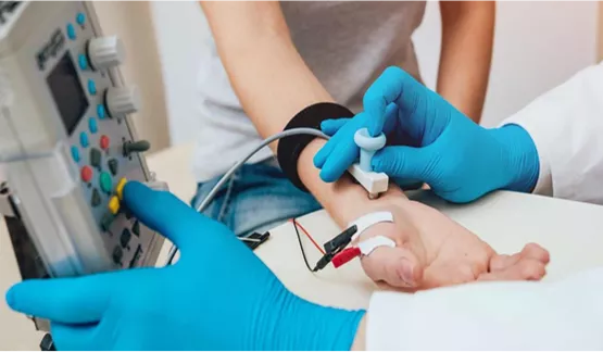
•
In an EMG procedure, small
needles called electrodes
are inserted into a patient’s
muscles in order to
measure the electrical
activity.
What does an EMG measure?
●
Your muscles move when (electrical) nerve signals from your
brain tell them to work. An EMG is used to measure how well your
muscles respond to these signals.
○
The electrodes in an EMG device are used to measure the electrical activity
caused at different levels of muscle contraction:
rest
,
slight contraction
, and
forceful contraction
.
○
This activity is then displayed on a monitor called an
oscilloscope
in the form
of
waves
.
https://www.amazon.com/Siglent-Technologies-
SDS1202X-Oscilloscope-Channels/dp/B07PVZMV16
EMG Waves
●
The size and shape of a wave, which represents an
action
potential
, gives information about how the muscle responds to
nerve stimulation.
○
A healthy muscle will show no electrical activity during rest, meaning it will not
show any signs of an action potential. It will only show electric activity when the
muscle moves or contracts.
○
When you contract your muscle more forcefully, more and more muscle fibers
are activated, making more action potentials.
EMG Waves
●
Here, you can see how the activity of a normal muscle is
represented.
○
Each blip, or wave, represents the firing of an action potential by a
motor
neuron
.
https://neurology.mhmedical.com/data/books/1844/demyer8_ch7_f021.png
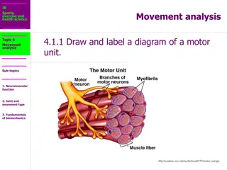
What is a Motor Neuron?
●
A
motor neuron
is a neuron that carries information from the
central nervous system to the muscles throughout our bodies
○
They are specialized cells which enable us to move by relaying information from
our brains to our muscles in the form of electric signals called
action potentials
,
such as when to contract and move.
https://smanewstoday.com/2017/09/12/sam-
research-may-get-boost-from-study-using-skin-
cells-to-create-human-motor-neurons/
Motor Neurons
●
There are two primary types of
motor neurons:
●
Upper motor neurons
,
originating
in a region of our brains called the
motor cortex, send electrical
signals (action potentials) to
lower
motor neurons.
●
Lower motor neurons
are
attached to our muscles and act as
a direct messengers to tell them to
start contracting.
https://impremedia.net/diagram-of-motor-neurone
/
EMG and Motor Neurons
●
These electrical signals, generated in motor neurons in the brain and sent
out to our muscles, are what EMG devices measure and record in order to
monitor the activity of muscles.
●
With the help of EMG, scientists are beginning to discover new
information about how movement is wired in our brain and bodies, and
how it can help detect neuromuscular disorders.
Electromyography (EMG) is an electrodiagnostic technique used to evaluate muscle health by measuring electrical activity. EMG measures muscle response to nerve signals, displayed as waves on an oscilloscope. Motor neurons transmit signals from the brain to muscles, enabling movement. Upper motor neurons originate in the motor cortex, while lower motor neurons directly command muscle contraction. EMG waves represent muscle activity, with each wave indicating an action potential fired by a motor neuron.

Download Presentation

Please find below an Image/Link to download the presentation.
The content on the website is provided AS IS for your information and personal use only. It may not be sold, licensed, or shared on other websites without obtaining consent from the author. Download presentation by click this link. If you encounter any issues during the download, it is possible that the publisher has removed the file from their server.
E N D
Presentation Transcript
What is an EMG? EMG stands for electromyography, which is an electrodiagnostic technique for evaluating and recording the electrical activity produced by skeletal muscles. We use them in order to assess the health of muscles and the nerve cells that help control them. In an EMG procedure, small needles called electrodes are inserted into a patient s muscles in order to measure the electrical activity. https://www.hopkinsmedicine.org/health/treatment-tests-and- therapies/electromyography-emg
What does an EMG measure? Your muscles move when (electrical) nerve signals from your brain tell them to work. An EMG is used to measure how well your muscles respond to these signals. The electrodes in an EMG device are used to measure the electrical activity caused at different levels of muscle contraction: rest, slight contraction, and forceful contraction. This activity is then displayed on a monitor called an oscilloscope in the form of waves. https://www.amazon.com/Siglent-Technologies- SDS1202X-Oscilloscope-Channels/dp/B07PVZMV16
EMG Waves The size and shape of a wave, which represents an action potential, gives information about how the muscle responds to nerve stimulation. A healthy muscle will show no electrical activity during rest, meaning it will not show any signs of an action potential. It will only show electric activity when the muscle moves or contracts. When you contract your muscle more forcefully, more and more muscle fibers are activated, making more action potentials.
EMG Waves Here, you can see how the activity of a normal muscle is represented. Each blip, or wave, represents the firing of an action potential by a motor neuron. https://neurology.mhmedical.com/data/books/1844/demyer8_ch7_f021.png
What is a Motor Neuron? A motor neuron is a neuron that carries information from the central nervous system to the muscles throughout our bodies They are specialized cells which enable us to move by relaying information from our brains to our muscles in the form of electric signals called action potentials, such as when to contract and move. https://smanewstoday.com/2017/09/12/sam- research-may-get-boost-from-study-using-skin- cells-to-create-human-motor-neurons/
Motor Neurons There are two primary types of motor neurons: Upper motor neurons, originating in a region of our brains called the motor cortex, send electrical signals (action potentials) to lower motor neurons. Lower motor neurons are attached to our muscles and act as a direct messengers to tell them to start contracting. https://impremedia.net/diagram-of-motor-neurone/
EMG and Motor Neurons These electrical signals, generated in motor neurons in the brain and sent out to our muscles, are what EMG devices measure and record in order to monitor the activity of muscles. With the help of EMG, scientists are beginning to discover new information about how movement is wired in our brain and bodies, and how it can help detect neuromuscular disorders.