Understanding the Respiratory System: An Overview
The respiratory system comprises the upper and lower respiratory tracts, responsible for air movement and gas exchange. It relies on the cardiovascular system for proper blood flow. The anatomy involves structures in the upper and lower tracts, with common symptoms like dyspnea and cough indicating various pulmonary and cardiac conditions. This comprehensive guide delves into the functions and structures of the respiratory system prepared by Ali Malik Tiryag.

Download Presentation

Please find below an Image/Link to download the presentation.
The content on the website is provided AS IS for your information and personal use only. It may not be sold, licensed, or shared on other websites without obtaining consent from the author. Download presentation by click this link. If you encounter any issues during the download, it is possible that the publisher has removed the file from their server.
E N D
Presentation Transcript
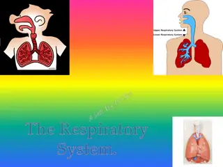
Respiratory System Prepared by Ali Malik Tiryag
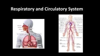
Anatomic and Physiologic Overview The respiratory system is composed of the upper and lower respiratory tracts. Together, the two tracts are responsible for ventilation (movement of air in and out of the airways). The upper respiratory tract, known as the upper airway, warms and filters inspired air so that the lower respiratory tract (the lungs) can accomplish gas exchange or diffusion. Gas exchange involves delivering oxygen to the tissues through the bloodstream and expelling waste gases, such as carbon dioxide, during expiration. The respiratory system depends on the cardiovascular system for perfusion, or blood flow through the pulmonary system
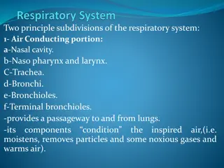
Anatomy of the Respiratory System Upper Respiratory Tract Upper airway structures consist of the nose; paranasal sinuses; pharynx, tonsils, and adenoids; larynx; and trachea. Lower Respiratory Tract The lower respiratory tract consists of the bronchi, bronchioles, alveolar ducts, and alveoli. Except for the right and left mainstem bronchi, all lower airway structures are found within the lungs. The right lung is divided into 3 lobes (upper, middle, and lower) and the left lung into 2 lobes (upper and lower)
Common Symptoms 1- Dyspnea Dyspnea (subjective feeling of difficult, breathlessness, shortness of breath) is a multidimensional symptom common to many pulmonary and cardiac disorders. Dyspnea may also be associated with allergic reactions, anemia, neurologic or neuromuscular disorders, trauma, and advanced disease, and is common at the end of life. Dyspnea can also occur after exercise in people without disease. In general, acute diseases of the lungs produce a more severe grade of dyspnea than do chronic diseases. Sudden dyspnea in a healthy person may indicate pneumothorax (air in the pleural cavity), acute respiratory obstruction, allergic reaction, or myocardial infarction.
In immobilized patients, sudden dyspnea may denote pulmonary embolism (PE). Dyspnea and tachypnea (abnormally rapid respirations) accompanied by progressive hypoxemia (low blood oxygen level) in a person who has recently experienced lung trauma, shock, cardiopulmonary bypass, or multiple blood transfusions may signal ARDS. Orthopnea (shortness of breath when lying flat, relieved by sitting or standing) may be found in patients with heart disease and occasionally in patients with chronic obstructive pulmonary disease (COPD); dyspnea with an expiratory wheeze occurs with COPD.
2- Cough Cough is a reflex that protects the lungs from the accumulation of secretions or the inhalation of foreign bodies. Its presence or absence can be a diagnostic clue because some disorders cause coughing and others suppress it. The cough reflex may be impaired by weakness or paralysis of the respiratory muscles, prolonged inactivity, the presence of a nasogastric tube, or depressed function of the brain s medullary centers (e.g., anesthesia, brain disorders). Cough results from irritation or inflammation of the mucous membranes anywhere in the respiratory tract and is associated with multiple pulmonary disorders. Mucus, pus, blood, or an airborne irritant, such as smoke or a gas, may stimulate the cough reflex.
Common causes of cough include asthma, gastrointestinal reflux disease, infection, and side effects of medications, such as angiotensin-converting enzyme (ACE) inhibitors. To help determine the cause of the cough, the nurse inquires about the onset and time of coughing. Coughing at night may indicate the onset of left-sided heart failure or bronchial asthma. A cough in the morning with sputum production may indicate bronchitis. A cough that worsens when the patient is supine suggests postnasal drip (rhinosinusitis). Coughing after food intake may indicate aspiration of material into the tracheobronchial tree. A cough of recent onset is usually from an acute infection.
The nurse assesses the character of the cough and associated symptoms. A dry, irritative cough is characteristic of an upper respiratory tract infection of viral origin, or it may be a side effect of ACE inhibitor therapy. An irritative, high-pitched cough can be caused by laryngotracheitis. A brassy cough is the result of a tracheal lesion, and a severe or changing cough may indicate bronchogenic carcinoma. Pleuritic chest pain that accompanies coughing may indicate pleural or chest wall (musculoskeletal) involvement. Violent coughing causes bronchial spasm, obstruction, and further irritation of the bronchi and may result in syncope (fainting).
3- Sputum Production Sputum production is the reaction of the lungs to any constantly recurring irritant and often results from persistent coughing. It may also be associated with a nasal discharge. The nature of the sputum is often indicative of its cause. A profuse amount of purulent sputum (thick and yellow, green, or rust colored) or a change in color of the sputum is a common sign of a bacterial infection. Thin, mucoid sputum frequently results from viral bronchitis. A gradual increase of sputum over time may occur with chronic bronchitis or bronchiectasis. Pink-tinged mucoid sputum suggests a lung tumor. Profuse, frothy, pink material, often welling up into the throat, may indicate pulmonary edema. Foul-smelling sputum and bad breath point to the presence of a lung abscess, bronchiectasis, or an infection caused by fusospirochetal or other anaerobic organisms.
4- Chest Pain Chest pain or discomfort may be associated with pulmonary, cardiac, gastrointestinal, or musculoskeletal disease or anxiety. Chest pain associated with pulmonary conditions may be sharp, stabbing, and intermittent, or it may be dull, aching, and persistent. The pain usually is felt on the side where the pathologic process is located, although it may be referred elsewhere for example, to the neck, back, or abdomen. Chest pain may occur with pneumonia, pulmonary infarction, or pleurisy, or as a late symptom of bronchogenic carcinoma. In carcinoma, the pain may be dull and persistent because the cancer has invaded the chest wall, mediastinum, or spine.
the parietal pleura have a rich supply of sensory nerves that are stimulated by inflammation and stretching of the membrane. Pleuritic pain from irritation of the parietal pleura is sharp and seems to catch on inspiration; patients often describe it as being like the stabbing of a knife. Patients are more comfortable when they lay on the affected side because this position splints the chest wall, limits expansion and contraction of the lung, and reduces the friction between the injured or diseased pleurae on that side. Pain associated with cough may be reduced manually by splinting the rib cage.
5- Wheezing Wheezing is a high-pitched, musical sound heard on either expiration (asthma) or inspiration (bronchitis). It is often the major finding in a patient with bronchoconstriction or airway narrowing. Rhonchi are low-pitched continuous sounds heard over the lungs in partial airway obstruction. Depending on their location and severity, these sounds may be heard with or without a stethoscope. 6- Hemoptysis Hemoptysis is the expectoration of blood from the respiratory tract. It can present as small to moderate blood-stained sputum to a large hemorrhage and always warrants further investigation. The onset of hemoptysis is usually sudden, and it may be intermittent or continuous.
The most common causes are: Pulmonary infection, Carcinoma of the lung, Abnormalities of the heart or blood vessels, Pulmonary artery or vein abnormalities, PE or infarction. Potential sources of bleeding include the gums, nasopharynx, lungs, or stomach. Bloody sputum from the nose or the nasopharynx is usually preceded by considerable sniffing, with blood possibly appearing in the nose. Blood from the lung is usually bright red, frothy, and mixed with sputum. Initial symptoms include a tickling sensation in the throat, a salty taste, a burning or bubbling sensation in the chest, and perhaps chest pain, in which case, the patient tends to splint the bleeding side. This blood has an alkaline pH (greater than 7). Blood from the stomach is vomited rather than expectorated, may be mixed with food, and is usually much darker and often referred to as coffee ground emesis. This blood has an acid pH (less than 7).
Diagnostic Tests 1-Pulmonary Function Tests Pulmonary function tests are noninvasive tests that show how well the lungs are working. The tests measure lung volume, capacity, rates of flow, and gas exchange. This information can help your healthcare provider diagnose and decide the treatment of certain lung disorders. 2-Arterial Blood Gas Studies Arterial blood gas (ABG) studies aid in assessing the ability of the lungs to provide adequate oxygen and remove carbon dioxide, which reflects ventilation, and the ability of the kidneys to reabsorb or excrete bicarbonate ions to maintain normal body pH, which reflects metabolic states.
PH 7.35 7.45 PCO2 35 45 mmHg PO2 75 100 mmHg HCO3 22 26 mEq/L SaO2 Greater than 95%
3-Pulse Oximetry Pulse oximetry, or SpO2, is a noninvasive method of continuously monitoring the oxygen saturation of hemoglobin (SaO2). A probe or sensor is attached to the fingertip, forehead, earlobe, or bridge of the nose. Normal SpO2 values are more than 95%. Values less than 90% indicate that the tissues are not receiving enough oxygen. 4- Cultures Throat, nasal, and nasopharyngeal cultures can identify pathogens responsible for respiratory infections, such as pharyngitis. Throat cultures are performed in adults with severe or ongoing sore throats accompanied by fever and lymph node enlargement and are most useful in detecting streptococcal infection. Other sources of infection, such as Staphylococcus aureus or Influenza, are detected via nasal or nasopharyngeal cultures. Ideally, all cultures should be obtained prior to the initiation of antibiotic therapy. Results usually take between 48 and 72 hours.
5- Sputum Studies Sputum is obtained for analysis to identify pathogenic organisms and to determine whether malignant cells are present. Periodic sputum examinations may be necessary for patients receiving antibiotics, corticosteroids, and immunosuppressive medications for prolonged periods because these agents are associated with opportunistic infections. Sputum samples ideally are obtained early in the morning before the patient has had anything to eat or drink. 6- Chest X-Ray Normal pulmonary tissue is radiolucent because it consists mostly of air and gases; therefore, densities produced by fluid, tumors, foreign bodies, and other pathologic conditions can be detected by x-ray examination.
7- Computerized tomography (CT) or positron emission tomography (PET) scans These are more advanced imaging tests that can be used to find problems that an X-ray might not until they re further along, like cancer. A CT scan is a series of X-rays taken from different angles that are put together to make a more complete picture. A PET scan uses a special dye that lets your doctor see parts of your body more clearly. 8- Magnetic Resonance Imaging MRI is similar to a CT scan except that magnetic fields and radiofrequency signals are used instead of radiation. MRI is able to better distinguish between normal and abnormal tissues than CT and, therefore, yields a much more detailed diagnostic image. MRI is used to characterize pulmonary nodules; to help stage bronchogenic carcinoma (assessment of chest wall invasion); and to evaluate inflammatory activity in interstitial lung disease, acute PE, and chronic thrombolytic pulmonary hypertension.
9- Pulmonary Angiography Pulmonary angiography is used to investigate congenital abnormalities of the pulmonary vascular tree, and less frequently PE, when less invasive tests are inconclusive, but there is a high clinical suspicion for PE. To visualize the pulmonary vessels, a radiopaque agent is injected through a catheter, which has been initially inserted into a vein (e.g., jugular, subclavian, brachial, or femoral vein) and then threaded into the pulmonary artery.


 or positron emission")