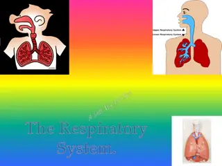
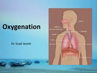
Understanding the Respiratory System and Its Functions
The respiratory system is vital for gas exchange in the body, involving processes like pulmonary ventilation, external and internal respiration. It includes organs like the nose, pharynx, larynx, and trachea, which help in conducting air to the alveoli. This system facilitates the exchange of oxygen and carbon dioxide between the blood and cells, supporting cellular respiration. Understanding the anatomy and functions of the respiratory system is crucial for overall health and well-being.

Download Presentation

Please find below an Image/Link to download the presentation.
The content on the website is provided AS IS for your information and personal use only. It may not be sold, licensed, or shared on other websites without obtaining consent from the author. Download presentation by click this link. If you encounter any issues during the download, it is possible that the publisher has removed the file from their server.
E N D
Presentation Transcript
Respiratory System: Overview Figure 17-2 b: Anatomy Summary
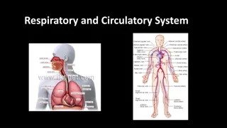
Respiration Includes Pulmonary ventilation Air moves in and out of lungs Continuous replacement of gases in alveoli (air sacs) External respiration Gas exchange between blood and air at alveoli O2 (oxygen) in air diffuses into blood CO2 (carbon dioxide) in blood diffuses into air Transport of respiratory gases Between the lungs and the cells of the body Performed by the cardiovascular system Blood is the transporting fluid Internal respiration Gas exchange in capillaries between blood and tissue cells O2 in blood diffuses into tissues CO2 waste in tissues diffuses into blood 3
Functions of the Respiratory System: Overview Exchange O2 Air to blood Blood to cells Exchange CO2 Cells to blood Blood to air Figure 17-1: Overview of external and cellular respiration
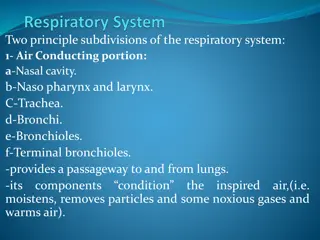
The Airways: Conduction of Air from Outside to Alveoli Filter, warm & moisten air Nose, (mouth), pharynx, larynx, trachea, bronchi & bronchioles Huge increase in cross sectional area Figure 17-4: Branching of the airways
Nose Provides airway Moistens and warms air Filters air External nose 6
The Pharynx (throat) Houses tonsils (they respond to inhaled antigens) Uvula closes off nasopharynx during swallowing so food doesn t go into nose Epiglottis posterior to the tongue: keeps food out of airway serve as common passageway for food and air * * 7
The Larynx (voicebox) Three functions: 1. Produces vocalizations (speech) 2. Provides an open airway (breathing) 3. Switching mechanism to route air and food into proper channels Closed during swallowing Open during breathing 8
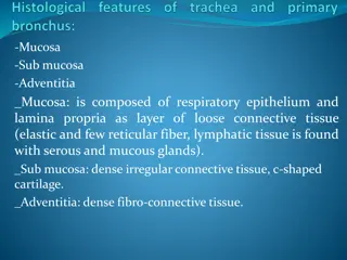
Trachea (the windpipe) Descends: larynx through neck Divides in thorax into two main (primary) bronchi 16-20 C-shaped rings of hyaline cartilage joined by fibroelastic connective tissue Flexible for bending but stays open despite pressure changes during breathing 9
Wider, shorter, and more vertical than the left trachea Right Primary Bronchus Left primary bronchus Both primary bronchi have the same anatomic structure as the trachea.
Bronchi The primary bronchi divide to form SECONDARY BRONCHI There is one secondary bronchus for each lobe of the lungs. There are 2 lobes on the left lung. There are 3 lobes on the right lung. These also have the same anatomy as the trachea.
Bronchi, continued The secondary bronchi branch to form TERTIARY BRONCHI. They continue to branch. As they get smaller, they lose their cartilage. When they lose their cartilage, they are called BRONCHIOLES which are microscopic.
Lungs and Pleura Around each lung is a flattened sac of serous membrane called pleura Parietal pleura outer layer Visceral pleura directly on lung Pleural cavity slit-like potential space filled with pleural fluid Lungs can slide but separation from pleura is resisted (like film between 2 plates of glass) Lungs cling to thoracic wall and are forced to expand and recoil as volume of thoracic cavity changes during breathing 13
Right lung: 3 lobes Upper lobe Middle lobe Lower lobe Left lung: 2 lobes Upper lobe Lower lobe Abbreviations in medicine: e.g. RLL pneumonia Horizontal fissure Oblique fissure Oblique fissure Each lobe is served by a lobar (secondary) bronchus 14


")
")
")