Comprehensive Overview of PACES Respiratory Examination and Management

Delve into the detailed outline, types of cases, examination tips, presentation wishlist, investigations, and management strategies for PACES respiratory cases. Learn about common conditions like ILD, bronchiectasis, and COPD, along with approaches and specific considerations in the examination process. Uncover the key aspects of patient presentation, diagnostic investigations, and multidisciplinary management approaches for respiratory disorders.

Download Presentation

Please find below an Image/Link to download the presentation.
The content on the website is provided AS IS for your information and personal use only. It may not be sold, licensed, or shared on other websites without obtaining consent from the author. Download presentation by click this link. If you encounter any issues during the download, it is possible that the publisher has removed the file from their server.
E N D
Presentation Transcript
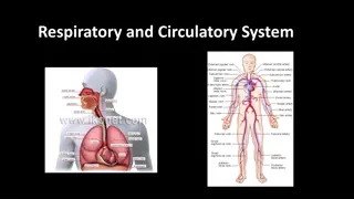
PACES Respiratory Wang Zhemin
Outline Types of Cases Approach Examination Tips Presentation Investigations Management Specific Conditions
Types of Cases Commonest ILD Bronchiectasis Pneumonectomy/Lobectomy Others Fibrothorax/Collapse/Consolidation COPD Pleural Effusion Pulmonary HTN
Approach Approach No Scar Scar Trachea Deviated: Pneumonectomy/ Upper Lobectomy ILD Trachea Central: Pneumonectomy/ Lobectomy Bronchiectasis Others
Examination Tips Hunt for the scar it can be pretty faint and well healed! Inspect for any autoimmune stigmata and RT marks Inspect carefully for symmetry when patient inspires Consider dextrocardia for Kartegener s If there isn t a scar, the money is often times at the back so ration your time wisely!
Presentation Wishlist: Complete my examination by reviewing patient s temperature charts, perform a bedside pulse oximetry, examine the content s of the patient s sputum mug and take a drug/occupational history (especially if ILD) If confident, cant a state diagnosis upfront Evidence by Cardinal features Comment on chest expansion, percussion, breath sounds (symmetry, well heard/reduced, adventitious), vocal resonance Comment on apex beat and trachea Complications (CPPRR): Cor pulmonale, Polycythemia, Pulm HTN, Respiratory failure, Respiratory Distress Etiology
Investigations Plain chest radiograph HRCT Pulmonary function test Invasive: Bronchoscopy (with BAL, TBLB), percutaneous biopsy
Management Multidisciplinary, involving respiratory physician and allied health staff, anchored in patient education Smoking cessation, Vaccination (Influenza, Pneumococcal), Chest Physiotherapy Symptomatic relief: Mucolytics, bronchodilators Treatment of infective exacerbations with antibiotics Treatment of underlying disease LTOT (at least 18 hours) PaO2</=55mmHg or SpO2 </=88% PaO2</=59mmgHg or SpO2 </=89% if 1) Cor pulmonale 2) RH failure 3) Erythrocytosis Surgical: Pneumonectomy/lobectomy (usually for complications), Lung transplant
ILD Features: Fine end inspiratory crepitations that do not change with coughing, dry cough, clubbing Causes Idiopathic Rheum: Psoriasis, Ank Spond, Systemic Sclerosis, DM/PM, RA Occupational (asbestosis, silicosis) Drugs (amiodarone, bleomycin, MTX, isoniazid) and Radiotherapy Apical vs Basal Apical: Rheum (Ankylosing spondylitis, Psoriasis, Sarcoidosis), Most pneumoconiosis except asbestosis (silicosis, coal worker pneumoconiosis, berrylosis), RT, TB, ABPA Basal: Idiopathic, Most rheumatological disorders (scleroderma, dermatomyositis, RA), asbestosis, drug induced (amiodarone, bleomycin, MTX, isoniazid)
ILD Investigations Plain Chest radiograph: Reticular-nodular shadowing High Resolution Computed Tomography: Ground glass changes, honey combing Pulmonary Function Test: Restrictive picture (FEV1/FVC > 70%, FVC < 80%, TLC < 80%) with reduced transfer factor (DLCO < 80%) Lung Biopsy Etiological Evaluation: Drug and occupational history RA: RF, anti-cyclic citrullinated peptide antibody SLE: ANA, anti-dsDNA, complements DM: Anti-Jo1 for antisynthetase Systemic sclerosis: Anti-centromere antibody (localized), anti-topoisomerase (ILD), anti-RNA polymerase (renal crisis) Complications: Echocardiogram (evaluate for pulmonary HTN PASP > 25) Treatment General treatment (as above), stop offending drug/occupational exposure, immunosuppression (steroid, immunomodulators), consider Perfenidone (antifibrotic) /Nintedanib (TKI) for IPF
Bronchiectasis Features: Coarse pan/early inspiratory crepitations that change on coughing, chesty cough, clubbing, rmb to check for dextrocardia Causes: Congenital: Cystic fibrosis, Kartegener s Syndrome (dextrocardia, situs inversus, primary ciliary dyskinesia), Hypogammaglobulinemia Acquired: Focal: Extraluminal (lymph nodes), luminal (tumour, previous TB), intraluminal (mucus plugging) Diffused: Post infectious (bronchiolitis obliterans), Rheumatological disorders (RA, SLE, Sjogren s), Yellow Nail Syndrome (Yellow nails, pleural effusion, lymphedema), ABPA
Bronchiectasis Investigations Plain Chest Radiograph: Tram tracks, ring shadows High Resolution Computed Tomography: Signet ring sign, dilated bronchi Pulmonary Function Test: Often obstructive picture (FEV1/FVC < 70%) Etiological Evaluation: Sputum culture, ciliary motility testing, genetic testing and sweat test for CF Treatment: General treatment (as above), bronchodilators, treat underlying cause Complications: Recurrent infection, hemoptysis, empyema, disseminated infection
Pneumonectomy/Lobectomy Clinical Features Findings are a SPECTRUM! Tracheal Deviation: Pneumonectomy or Upper Lobectomy Lobectomy: Findings are generally normal because rest of lung fills up Pneumonectomy: Tracheal deviation, bronchial breath sounds adjacent to trachea, rest of lung reduced chest expansion, dullness to percussion, absent breath sounds, decreased vocal resonance Causes: Cancer (Early NSCLC), Abscess, Bronchiectasis with hemoptysis, Tuberculosis, Lung volume reduction surgery, Trauma
Pleural Effusion Light s Criteria (1 out of 3 exudative) Pleural protein : serum protein > 0.5 Pleural LDH : serum LDH > 0.6 Pleural LDH > 2/3 upper limit of normal serum LDH level Other pleural fluid tests: Microscopy: Cell count, cytology Microbiological: Gram stain, cultures, AFB Biochemistry: pH (<7.2 suggestive of empyema), glucose (low: malignancy, empyema, RA, TB), ADA, protein, LDH, haematocrit (haemothorax) Causes Transudative: Fluid overload (heart failure, renal failure), hypoalbuminemia (nephrotic syndrome, chronic liver disease, hypothyroidism), Exudative: Parapneumonic effusion, empyema, TB, malignancy, connected tissue disorders Others: Chylothorax, haemothorax