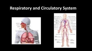
Understanding the Cardio-Respiratory System in GCSE PE
Explore the structure and function of the cardio-respiratory system in GCSE PE, including the pathway of air, gaseous exchange, cardiac cycle, and key terminologies. Learn how the respiratory system serves to deliver oxygen to muscles and remove carbon dioxide. Discover the mechanisms of breathing, the impact of exercise on breathing, and how the lungs and heart adapt during physical activity.

Download Presentation

Please find below an Image/Link to download the presentation.
The content on the website is provided AS IS for your information and personal use only. It may not be sold, licensed, or shared on other websites without obtaining consent from the author. Download presentation by click this link. If you encounter any issues during the download, it is possible that the publisher has removed the file from their server.
E N D
Presentation Transcript
Cirencester Kingshill School PE Department GCSE PE Chapter 1b The Structure and Function of the Cardio- Respiratory System At the end of three theory lessons you will know Pathway of air through the body Understand gaseous exchange Cardiac Cycle Key terminology such as Stroke Volume, Cardiac output
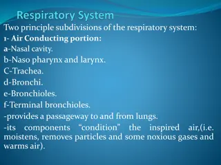
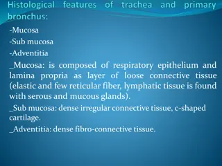
Functions What purpose does the Respiratory system serve? To get Oxygen from the air into the lungs, which is then transported to the required muscles via our blood To receive waste Carbon Dioxide, to exhale in the air.
Show me your H/W USE TEXT BOOK TO CHECK IF YOU HAVE LABELLED THE DIAGRAM CORRECTLY stick in book!
Mechanisms of breathing Inspiration Intercostal muscles pull ribs up and out When you breathe in: intercostal muscles between the ribs contract, pulling the chest walls up and out the diaphragm muscle below the lungs contracts and flattens, increasing the size of the chest the lungs increase in size, so the pressure inside them falls. This causes air to rush in through the nose or mouth. Diaphragm contracts and moves down next_btn_colour 4
Mechanisms of breathing Expiration When you breathe out: Ribs move in and down Intercostal muscles between the ribs relax so that the chest walls move in and down. The diaphragm muscle below the lungs relaxes and bulges up, reducing the size of the chest. The lungs decrease in size, so the pressure inside increases and air is pushed up the trachea and out through the nose or mouth. Diaphragm relaxes and bulges up next_btn_colour 5
Complete this table INTERCOSTALS/RIBS DIAPHRAGM LUNGS INSPIRATION EXPIRATION
Breathing during exercise During exercise the muscle cells use up more oxygen and produce increased amounts of carbon dioxide. Your lungs and heart have to work harder to supply the extra oxygen and remove the carbon dioxide. Your breathing rate increases and you breathe more deeply. Heart rate also increases in order to transport the oxygenated blood to the muscles. next_btn_colour 7
Breathing during exercise Muscle cell respiration increases more oxygen is used up and levels of CO2 rise. The brain detects increasing levels of CO2 a signal is sent to the lungs to increase breathing. Breathing rate and the volume of air in each breath increase. This means that more gaseous exchange takes place. The brain also tells the heart to beat faster so that more blood is pumped to the lungs for gaseous exchange. More oxygenated blood gets to the muscles and more CO2 is removed. next_btn_colour 8
H/W USE TEXT BOOK TO RESEARCH Lung Volumes Match the definitions and fill in the Spirograph. Due 19/10
LUNG VOLUMES Tidal volume is the amount you breathe in and out in one normal breath. Respiratory rate is how many breaths you take per minute. Minute volume is the volume of air you breathe in one minute. Vital capacity is the maximum volume of air you can breathe out after breathing in as much as you can. Residual volume is the amount of air left in your lungs after you have breathed out as hard as you can. next_btn_colour
Calculating minute volume Remember: Minute volume is the volume of air you breathe in one minute. You can calculate a person s minute volume by multiplying the volume of air they breathe in one breath, by their respiratory (breathing) rate. Question If you breathe 14 times in one minute (respiratory rate) and you breathe 0.5 litres in each breath, what is your minute volume? Answer: Minute volume = 14 0.5 litres = 7.0 litres next_btn_colour
Gas exchange at the alveoli The alveoli are bunches of tiny air sacks inside the lungs. Each individual sack is called an alveolus. When you breathe in, they fill with air. The alveoli are covered in tiny capillaries (blood vessels). Gases can pass through the thin walls of each alveolus and capillary, and into the blood stream. Gases can also pass from the blood stream, into the alveolus. next_btn_colour
Task: Draw and explain the function of the alveoli Equipment: White board Whiteboard pen Red and blue Red and blue Smarties 3 of each! (two different colours) 1. Draw Alveoli outline 2. Use pages 11 and 12 to annotate and describe 3. After 8 mins we will go through some answers be prepared to share. 4. Take a picture of your finished diagram and annotations. 5. Email to yourself and print out and stick in your book for next lesson!
Use page 11 and 12 to create Copy or bullet point this diagram further notes on this process. next_btn_colour
Exam-style questions 1. Describe the passage of oxygen from the nasal passages to the bloodstream. 2. David goes jogging once a week for 45 minutes. a) List two differences between the air that David inhales and the air that he exhales while jogging. b) What two substances are used by David s body cells to produce energy? What are the products of this reaction? David tries to increase his pace. He finds that he is forced to stop running and breathe hard for several minutes. c) Explain why David had to stop. d) How did breathing hard help him to recover? next_btn_colour
CHECK you have all the terminology written down!!! Page 13 EXTENSION work Complete the CHECK YOUR UNDERSTANDING QUESTIONS (page 12 and 13)
CIRCULATORY SYSTEM
Functions of the blood 1. Transporting substances around the body. These include oxygen, glucose, carbon dioxide, nutrients, water and waste products. 2. Controlling body temperature. 3. Protecting the body. Blood contains cells and anti-bodies that fight infection and clotting agents to stop bleeding.
The Blood Blood is the body s means of transporting substances around. It transports: oxygen from the lungs to the heart and then to the body s tissues carbon dioxide from the tissues to the heart and then to the lungs to be expired materials like hormones from one organ to another nutrients (especially glucose) and minerals from the intestines to the tissues waste products to the kidneys.
Blood Vessels There are three types of blood vessels, as shown in this magnified part of the circulatory system. blood to the heart blood from the heart vein artery carries blood away from the heart carries blood back into the heart carries blood to and from the body s cells Why are there different types of blood vessels?
Blood Vessels thick outer wall thick inner layer of muscle and elastic fibres narrow central tube (lumen) ARTERY thin outer wall thin inner layer of muscle and elastic fibres CAPILLARY wide central tube (lumen) VEIN wall only one cell thick
Veins When blood is flowing against gravity, or when a vein is squeezed by muscle action, there is a risk that blood will flow in the wrong direction. Veins have valves to prevent backflow. backflow prevented vein valve open vein valve closed blood to the heart The valves allow blood to flow in the correct direction but close if blood starts to flow in the wrong direction.
H/W USE TEXT BOOK TO RESEARCH The Cardiac Cycle fill in your sheet (use your previous heart homework) Due After half term 15/11
Cardiac system The four chambers of the heart have special names: An upper chamber is called an atrium (plural: atria). right atrium left atrium right ventricle left ventricle A lower chamber is called a ventricle. next_btn_colour
Cardiac system Here are some other important parts of the heart: The semi-lunar valves prevent expelled blood flowing back into the heart. The walls are made of cardiac muscle. Bicuspid (mitral) valve The wall dividing the left and right sides of the heart is called the septum. Tricuspid valve These two valves prevent blood flowing back into the atria from the ventricles. next_btn_colour
Cardiac Cycle The cardiac cycle is the sequence of events that occurs when the heart beats. As the heart beats, it circulates blood through pulmonary and systemic circuits of the body. In the diastole phase, the heart ventricles are relaxed and the heart fills with blood. In the systole phase, the ventricles contract and pump blood out of the heart and to arteries. One cardiac cycle is completed when the heart chambers fill with blood and blood is then pumped out of the heart.
The heart during exercise Each individual has a maximum heart rate the fastest that their heart is able to beat. Testing it properly is difficult and unpleasant, as it involves pushing your body to its absolute limit. However, maximum heart rate can be estimated using a simple formula: Maximum Heart Rate (MHR) = 220 age So, a 25 year-old would have a maximum heart rate of 220 25 = 195 bpm What would your maximum heart rate be? next_btn_colour
Heart rate, stroke volume and cardiac output The pulse rate is not the only way of measuring the heart. Stroke volume is the amount of blood pumped out of the left ventricle per beat. Cardiac output is the amount of blood pumped out of the left ventricle of the heart per minute. Cardiac output can be calculated by multiplying the stroke volume by the heart rate: cardiac output = stroke volume heart rate What is the cardiac output of someone with a heart rate of 60 bpm and stroke volume of 90 ml? next_btn_colour
The heart during exercise During exercise, the body uses up oxygen and nutrients at a much faster rate. To keep the body supplied with what it needs, the heart beats faster and with greater force. This means that the heart rate and stroke volume increase. What do you think happens to the cardiac output? Regular exercise causes changes to the heart. The heart gets larger The muscular wall become thicker and stronger Stroke volume at rest increases, leading to a lower resting heart rate. next_btn_colour
Can you remember all these keywords? Aorta Vena cava Pulmonary artery Pulmonary vein Pulmonary circulation Systemic circulation Oxygenated Deoxygenated Arteries Veins Capillaries Lumen Atria Ventricles Septum Tricuspid valve Bicuspid (mitral) valve Semi-lunar valve Heart rate Stroke volume Cardiac output Blood pressure Vasodilation Vasoconstriction Red blood cells White cells Platelets Plasma Blood doping
CHECK the terminology (page 16) EXTENSION work Complete the CHECK YOUR UNDERSTANDING QUESTIONS (page 17)