Understanding Nasal Cavity Histology and Respiratory Structures
The detailed information provided covers the histological structures of the nasal cavity, including the vestibule, respiratory mucosa, nasal septum, olfactory mucosa, and paranasal sinuses. It also delves into the microscopic structures of the wall of the trachea and primary bronchi. The respiratory system's components and divisions, from the nasal cavity to the alveoli, are explained thoroughly, aiding in a comprehensive understanding of the respiratory tract anatomy and function.

Download Presentation

Please find below an Image/Link to download the presentation.
The content on the website is provided AS IS for your information and personal use only. It may not be sold, licensed, or shared on other websites without obtaining consent from the author. Download presentation by click this link. If you encounter any issues during the download, it is possible that the publisher has removed the file from their server.
E N D
Presentation Transcript
Respiratory Block | Histology Color index: -Main text -important -female slides -male slides -Dr.note -Extra Upper Respiratory Tract Editing file Editing file
Objectives By the end of this lecture the student should be able to describe the microscopic structures of: Vestibule of the nasal cavity. Respiratory mucosa of the nasal cavity. Nasal septum. Olfactory mucosa of the nasal cavity. Mucosa of the paranasal sinuses. Larynx. - The microscopic structures of the wall of: Trachea. Primary or extra-pulmonary bronchi.
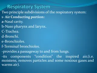
RESPIRATORY SYSTEM 1- Nasal cavity. 2- Nasopharynx. 3- Larynx. 4- Trachea. 5- Primary bronchi (extrapulmonary bronchi). 6- Intrapulmonary bronchi: - 2ry bronchi (lobar bronchi). - 3ry bronchi (segmental bronchi). 7- Primary bronchioles (preterminal bronchioles). 8- Terminal bronchioles. Conducting portion 1- Respiratory bronchioles. 2- Alveolar ducts. 3- Alveolar sacs. 4- Pulmonary alveoli. Respiratory portion
NASAL CAVITY (N.C.) N.B. The nasal septum divides the nasal cavity into two halves (right and left). Anterior portion Posterior portion (Vestibule of nasel cavity ) Lining: linked with the skin 1- Epidermis:(Keratinized stratified Squamous epithelium). 2- Dermis. Contents: 1- Vibrissae: stiff hairs. 2- Sebaceous glands. 3- Sweat glands. Wall: 1- Hyaline cartilage.(majority of respiratory tract) 2-Cancellous (spongy) bone. Respiratory region Olfactory region Will be explained in the next slides
RESPIRATORY REGION OF NASAL CAVITY MUCOSA (MUCOUS MEMBRANE): Pseudo-stratified ciliated columnar epithelium with goblet cells. Main types of cells: (all touch the basement membrane) 1- Ciliated columnar cells. 2- Goblet cells. 3- Basal cells: are stem cells. 4- DNES(diffuse neuroendocrine) cells: e.g. serotonin. Respiratory epithelium Main types of cells 1- Large arterial plexuses & venous sinuses (Highly vascularized C.T.) (dialited blood vessels to warm the air) 2- Many seromucous glands (acini). 3-Abundant lymphoid elements: Including occasional lymphoid nodules, plasma cells & mast cells. Lamina propria (sub-epithelial C.T.) Contains - epithelium+connective tissue in wet area is called mucous membrane - The only neuron that can renew from its stem cell is located in olfactory area
OLFACTORY REGION (AREA) OF NASAL CAVITY (OLFACTORY MUCOSA) site 1-Roof of nasal cavity. 2-Upper part of nasal septum. 3-over superior concha. Olfactory epithelium: Pseudo-stratified columnar epithelium. - Are bipolar neurons (dendrite+axon) - Dendrite has olfactory vesicle that has nonmotile cilia. (to increase surface area) - Axons are unmyelinated with Schwann-like cells. (nerve fibers supporting cells of neuron axon) - Axons will collect in the lamina propria to form bundles of nerve fibers. Bundles will collect to form the olfactory nerve. Olfactory cells(olfactory nerve cells) Olfactory epithelium -Are columnar cells. -function: Physical support and nourishment for olfactory cells. Sustentacular (supporting) cells Basal cells Pyramidal in shape, basal in position and act as stem cells. Lamina propria Highly (richly) vascularized loose C.T. Contents a) Bowman s glands (olfactory glands): are serous acini. b) Bundles of unmyelinated nerve fibers: Are axons of olfactory nerve cells+Schwann-like cells (glial cells). c) Rich vascular plexus. d) Numerous lymphoid elements (there s no goblet cells or mucus glands)
Vestibular folds: Are immovable. L/M: a- Respiratory epithelium. b- Lamina propria; Loose connective tissue .with seromucous glands, lymphoid elements & adipose cells. Lamina propria Extrinsic & intrinsic all are skeletal Vocal folds (cords), have: a- Epithelium: non keratinized stratified squamous. b- Lamina propria: connective tissue. containing bundles elastic fibers and skeletal muscle. -No lymphoid nodules, No seromucous glands. Larynx 1-hyaline cartilage e.g.thyroid 2-elastic cartilage e.g. epiglottis 1- Epithelium: (2 types) a- Respiratory epithelium: Pseudostratified ciliated columnar epithelium with goblet cells. b- Non keratinized stratified squamous epithelium: In: -Vocal folds. - Superior surface of epiglottis Epithelium
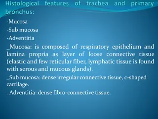
TRACHEA 1-Epithelium Respiratory epithelium mucosa 2-Lamina propria 3-Elastic lamina It is formed of elastic fibers. It separates lamina propria from submucosa. 1-Connective tissue 2-Numerous mucous & seromucous glands. 3-Lymphoid elements. Fibroelastic connective tissue C-shaped rings (12-16) of hyaline cartilage. Trachealis muscle (bundle of smooth muscle fibers) connects the 2 ends of each C-shaped (incomplete) rings of cartilage. submucosa Adventitia PARANASAL SINUSES Lining Clinical application 1- Respiratory epithelium. 2- Lamina propria. Sinusitis
Extrapulmonary Bronchus (1ry Bronchus) Generally have the same histological appearance as the trachea.
MCQs: Click the icon for more questions 1-Which of the following is not a part of conducting portion in respiratory system? A-larynx B-trachea C-Alveolar ducts D-Intrapulmonary bronchi 2-what type of cartilage is found in the wall of nasal cavity? C-fibrocartilage D-non A-hyaline cartilage 3-seromucous glands (acini). Is found in? B-elastic cartilage B-respiratory region of nasal cavity A-olfactory region of nasal cavity 4-DNES cells: e.g. serotonin. are in? D-larynx C-trachea A-olfactory region of nasal cavity 5-Vocal folds (Cords) are in ? B-respiratory region of nasal cavity D-larynx C-trachea A-pharynx B- paranasal sinuses C-trachea D-larynx 6-which of the following is not not found in trachea? 6-D 5-D 4-B D- skeletal muscle A- Fibroelastic C.T B- C-shaped hyaline cartilage C-smooth muscle 3-B 2-A 1-C
Respiratory block|Histology team 442 Team leaders: -Mashael Alsuliman. -Nawaf Almaiman Team members: -Nawaf Alturki -Leena Alabbad -Abdulkarim Salman -Lina Almohsen -Zyad Alodan 442Histology@gmail.com


")

 OF NASAL CAVITY (OLFACTORY MUCOSA)")


")