Veterinary Guide: Respiratory System Disorders in Horses
Respiratory system disorders in horses are crucial as they can impact the animals' performance and lead to economic losses for owners. Early detection and treatment are essential for ensuring the rapid return of athleticism in performance animals. This guide covers the diagnostic approach to respiratory disorders, including history taking and physical examination techniques like auscultation and percussion of the lung fields.

Download Presentation

Please find below an Image/Link to download the presentation.
The content on the website is provided AS IS for your information and personal use only. It may not be sold, licensed, or shared on other websites without obtaining consent from the author. Download presentation by click this link. If you encounter any issues during the download, it is possible that the publisher has removed the file from their server.
E N D
Presentation Transcript
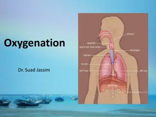
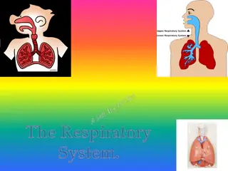
Disorders of the Respiratory System Dr. Bipin Kumar Assistant Professor MVSc VCM-603 Unit 2 Department of Veterinary Medicine Bihar Veterinary College, Patna (Bihar Animal Sciences University, Patna)
Disorders of the Respiratory System Second in importance only to those of the musculoskeletal system. Owners sustain major economic losses when respiratory diseases interrupt the training programs of horses or when horses must be retired because of lung damage sustained from respiratory disease. Thus early detection and treatment of respiratory problems is essential for the rapid return of athleticism to performance animals.
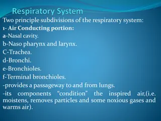
Diagnostic Approach to Respiratory Disorders HISTORY: The clinician should direct questions to the person most familiar with the performance and medical history of the horse. Age and Breed:The age and breed of the animal exhibiting respiratory-related signs may provide clues as to the problem. Congenital defects (nasal septal deviations, choanal atresia, subepiglottic cysts, and hypoplastic lungs) are typically evident at birth, whereas other conditions, such as chronic bacterial pneumonia (Rhodococcus equi), may not be evident until the foal is older (1 to 3 months of age). Environment One should ascertain the environment to which the horse was or presently is exposed. Prior Medical Problems Has the horse had a previous history of illness or trauma that might be related to the present complaint? Present Medical Problem
PHYSICAL EXAMINATION Before examining the horse, simply stepping back and evaluating the demeanor and mental status (alert or depressed), posture, and manner of movement of the horse is helpful. BREATHING PATTERNS Eupnea Tachypnea Hyperpnea Apnea Hypoventilation Hyperventilation Dyspnea
Auscultation of the Lung Fields The clinician should examine the horse during eupneic and hyperpneic (by use of a rebreathing bag) breathing patterns. Normal breath sounds are those produced by turbulent air movement through the tracheobronchial tree and vary in intensity and quality depending on the portion of the lung field auscultated. The vesicular sounds, over the middle and diaphragmatic lung lobes, are the quietest sounds; The bronchial sounds, over the trachea and the base of the lung, are the loudest. Adventitious lung sounds are abnormal sounds superimposed on the
Percussion of the Thorax One percusses by methodically tapping the intercostal spaces of the thorax using a plexor and pleximeter (foals) or a large spoon and neurologic hammer (adults) and evaluating the nature of the sound produced. Aerated tissues produce a resonant sound, whereas fluid-filled structures (bowel, heart, lung abscesses, consolidated lung) produce a dull sound. Radiography ULTRASONOGRAPHY Endoscopy Sinuscopy Computed Tomography
Sampling of Respiratory Tract Secretions Centesis of the Paranasal Sinuses One performs the technique aseptically using local anesthesia on the sedated horse, using a Steinmann pin for the initial puncture of the sinus. One enters the rostral maxillary sinus at a site 2.5 cm dorsal to the facial crest and 2.5 cm caudal to the infraorbital foramen. One enters the caudal maxillary sinus (which communicates with the frontal sinus) at a site 2.5 cm dorsal to the facial crest and 2.5 cm rostral to the medial canthus. The clinician should submit aspirates for cytologic examination and bacterial culture. Guttural Pouch Catheterization and Culture of the Exudate
Sampling of Tracheobronchial Secretions Several techniques have been advocated for obtaining tracheobronchial samples. Sedation of horses before bronchoalveolar lavage is recommended. Using a fiberoptic endoscope (permitting direct visualization of the lung segment to be lavaged) or using a thick-walled flexible tube with a cuffed end (which is passed blindly into the distal airways), the clinician instills 100 to 500 ml of physiologic saline solution within the pulmonary segment. One can retrieve 50% to 75% of the fluid and examine it cytologically. Before lavage, one may instill 20 to 40 ml of 2% lidocaine to desensitize the airways.