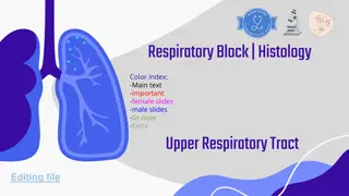
Respiratory Tract Anatomy Overview
The respiratory tract anatomy includes various structures like mucosa, submucosa, and adventitia in the trachea, bronchi, bronchioles, and terminal bronchioles. Each component has specific compositions and functions, ranging from respiratory epithelium to smooth muscle layers. The images provide a visual representation of these anatomical features, highlighting the differences in cartilage arrangement, epithelial types, and secretory cells present in different parts of the respiratory tract.

Download Presentation

Please find below an Image/Link to download the presentation.
The content on the website is provided AS IS for your information and personal use only. It may not be sold, licensed, or shared on other websites without obtaining consent from the author. Download presentation by click this link. If you encounter any issues during the download, it is possible that the publisher has removed the file from their server.
E N D
Presentation Transcript
-Mucosa -Sub mucosa -Adventitia _Mucosa: is composed of respiratory epithelium and lamina propria as layer of loose connective tissue (elastic and few reticular fiber, lymphatic tissue is found with serous and mucous glands). _Sub mucosa: dense irregular connective tissue, c-shaped cartilage. _Adventitia: dense fibro-connective tissue.
Intrapulmonary bronchi: (Secondary and tertiary bronchi) -The primary bronchi are divided into secondary bronchi in the lung which is covered by thick elastic serous membrane (the plura) composed of two layers parietal and visceral. -The internal structure of lung consists of branching system of bronchial tree. Secondary bronchus: -The mucosa is composed of respiratory epithelium and lamina propria contains connective tissue, serous and mucous glands.
-Irregular platesof cartilage instead of c-shaped cartilage. -smooth muscle layer as muscular is spirally arranged separated the lamina propria from sub mucosa, is rich with elastic fiber.
Trachea Trachea section H&E 10x objective ciliated pseudostratified columnar epithelium adventitia submucosa lumen hyaline cartilage adipose tissue
Bronchioles: -large bronchioles about 5mm or less in diameter. -the wall is lined by respiratory epithelium, no glands, no cartilage. -the epithelium becomes ciliated columnar with some goblet cells. -bundle of smooth muscle is increased run in spiral manner. 1-Terminal bronchioles: -Is distal part of bronchiole about 0.5 mm or less in diameter. -the epithelium is columnar epithelium ciliated and some non-ciliated, have no goblet cells,Epithelium gradually become mostly Clara cells with cuboidal rather than ciliated epithelium as the terminal bronchioles near the respiratory bronchioles.
-Clara cells, secretory cell, dome-shaped, secret surfactants that decrease surface tension of fluid that moistens and protect the surface of the bronchiolar lining. -lamina propria composed of elastic fiber, the smooth muscles, thick bundles one or two layers spirally arranged, so the mucosa show longitudinal folding.
2-Respiratory bronchioles: -are lined by simple cuboidal cells, some of them are ciliated and the wall is interrupted by simple squamous epithelium (the wall of alveoli). -the muscle and elastic fiber are found.
Alveolar duct: Has thin discontinuous lined by simple squamous epithelium supported by fibro elastic connective tissue. Alveolar sacs: Are formed the opening of several alveoli, attached to the ducts. Lung alveoli (pneumocytes): 200 million in a normal lung Total area-75 square meters Total capillary surface area available for exchange- 125square meters Are spongy and form the parenchyma of lung. Sac like evagination present at the terminal end of the bronchial tree.
Alveoli are separated by Interalveolar septum lying between thin epithelial lining of two neighboring alveoli. Interalveolar septum contains a network of capillaries supported by reticular and elastic fibers, occasionally fibroblasts, macrophages and mast cells. Septum contains pores(ALVEOLAR PORES OF KOHN) help in passage of air from one alveolus to another, thus equalizing Pressure in the alveoli. Elastic fibers-enable the alveoli to expand during inspiration and passively contract during expiration. Reticular fibers support and prevent over distention of the alveoli.
_Cells in Alveoli: Type I Pneumocytes Type II Pneumocytes Macrophages or Dust cells -Alveolar Type I cells They form about 45% of alveolar cells population, are lined by an thin simple squamous epithelium, their ends are connected by tight junction to prevent leakage of tissue fluid in to the alveolar lumen. -Alveolar Type II cells -They are roughly cuboidal with many microvilli on their free surface.
-synthesize and secret a surfactant, it reduces surface tension across the squamous epithelium of the alveoli in the lungs. -prevent collapse of alveoli during expiration. -regenerate themselves and replace the damaged type pneumocytes. -Alveolar macrophages-dust cells- usually free in the alveolar lumen, also in tissues between alveoli. Derived from Monocytes and are part mononuclear phagocytic system. Either seen in the septa or alveoli.
Alveoli Respiratory bronchiole cross section H&E 10x objective alveolar wall alveolar sac
Cytoplasm contains phagocytosed inhaled carbon and dust particles. Inhaled carbon and dust particles are passed on to them from pneumocyte I through pinocytic vesicles. Migrate from septum to alveolar surface and are carried to the pharynx through sputum. Main function is to clean the alveoli of invading microorganisms and inhaled particulate matter by phagocytosis.