Embryology and Pathogenesis of Spinal Dysraphism
Gastrulation, the formation of germ layers, mesoderm, and endoderm, in the embryo occurs during the third week of gestation with the appearance of the primitive streak. Cells migrate to form endoderm and mesoderm while the ectoderm remains. Neural induction is regulated by factors like fibroblast growth factor and bone morphogenetic protein 4, influencing the development of neural tissues. Molecular mechanisms involving chordin, noggin, and follistatin inactivate BMP4 and play vital roles in neural tissue induction. Retinoic acid and signaling pathways like WNT3a and FGF are crucial for hindbrain and spinal cord development.

Download Presentation

Please find below an Image/Link to download the presentation.
The content on the website is provided AS IS for your information and personal use only. It may not be sold, licensed, or shared on other websites without obtaining consent from the author. Download presentation by click this link. If you encounter any issues during the download, it is possible that the publisher has removed the file from their server.
E N D
Presentation Transcript
SPINAL DYSRAPHISM EMBRYOLOGY AND PATHOGENESIS
GASTRULATION Gastrulation is the process of establishment of the three germ layers, mesoderm and endoderm, in the embryo. namely, the ectoderm, Gastrulation occurs in the third week of gestation which begins with the appearance of the primitive streak, which has the primitive node at its cephalic end.
In the region of the node and streak, the epiblast cells move inward, i.e., invaginate, to form the endoderm and the mesoderm The cells that do not migrate through the streak but remain in the epiblast from the ectoderm
ECTODERMAL GERM ECTODERMAL GERM LAYER LAYER At the beginning of the 3rd week of development, the ectodermal layer has the shape of a disc that is broader in the cephalic than in the caudal region
ECTODERMAL GERM LAYER Appearance of the notochord and the precordal mesoderm induces the overlying ectoderm to thicken and form the neural plate Cells of the plate make up the neuroectoderm, and their induction represents the initial event in the process of neurulation
MOLECULAR REGULATION OF NEURAL INDUCTION Induction of neural plate is caused by the up regulation of fibroblast growth factor (FGF) with the inhibition of bone morphogenetic protein 4 (BMP4) FGF signaling promotes a neural pathway by an unknown mechanism while transcription and up regulates the expression of chordin and noggin, which inhibit BMP activity it represses BMP If BMP 4 is absent or is inactivated, the ectoderm becomes neutralized
MOLECULAR REGULATION OF NEURAL INDUCTION Secretion of chordin, noggin and folistatin inactivates BMP4. These neural inducers induce only forebrain and midbrain types of tissues, and the induction of the caudal neural plate structures ( hindbrain and spinal cord) depends on WNT3a and FGF. Also, retinoic acid plays a role in the organization of cranial to caudal axis.
NEURULATION The elongated neural plate gradually expands towards the primitive streak. The lateral edges of the neural plate become more elevated by the end of the third week to form neural folds, and the depressed mid region forms the neural groove.
NEURULATION The fusion begins in the cervical region and proceeds cranially and caudally. As a result, the neural tube is formed. Until fusion is complete, the cephalic and the caudal ends of the neural tube communicate with the amniotic cavity by way of the cranial neuropores, respectively. and caudal
NEURULATION The cranial neuropore closes at approximately day 25, while the posterior neuropore closes on day 27. Thus, at the completion of neurulation , the CNS is represented by the spinal cord, a closed tubular structure with a narrow caudal portion, and a broader cephalic portion, the brain vesicles.
SPINAL CORD Neuro epithelial, mantle and Marginal Layers Once neuroepithelial cells give rise to the primitive nerve cells, namely neuroblasts. They form the mantle layer which forms the grey matter of the spinal cord. the neural tube closes, the The outermost layer of the spinal cord, the marginal layer, contains nerve fibres arising from neuroblasts in the mantle layer. As a result of myelination of the nerve fibres, this layer has a white appearance and is therefore called the white matter of the spinal cord.
POSITIONAL CHANGES OF THE SPINAL CORD In the 3rd month of development, the spinal cord extends the entire length of the embryo, and spinal nerves intervertebral foramina at their level of origin. pass through the With increasing age, the vertebral column and dura lengthen more fate than the neural tube, and the terminal end of the cord shifts to a higher level. At birth, this level is at L3.
POSITIONAL CHANGES OF THE SPINAL CORD The disproportionate growth results in oblique direction of the spinal nerves from their segment of origin in the cord to the corresponding level in the vertebral column.
POSITIONAL CHANGES OF THE SPINAL CORD In the adult, the spinal cord terminates at the lower border of L1 or L2, and the dural sac and the subarachnoid space extend to S2.
POSITIONAL CHANGES OF THE SPINAL CORD Below L2 to L3, a threadlike extension of the pia mater forms the filum terminale, which is attached to the periosteum of the 1st coccygeal vertebra. Nerve fibres below the terminal end of the cord collectively constitute the cauda equina.
SPINAL DYSRAPHISM Most of the defects of the spinal cord result from abnormal closure of the neural folds in the 3rd and 4th weeks of development. Spinal dysraphism refers to all forms of spina bifida. Spina bifida is a general term for neural tube defects affecting the spinal region.
SPINAL DYSRAPHISM - PATHOGENESIS HISTORY Failure of closure of neural tube. First described by von Recklinghausen in 1886. Excessive overgrowth of neural tissue in the region of a spinal or cranial defect. Suggested by Patten in 1953. Padget proposed abnormal neuroschisis . The hydromyelic theory was proposed by Morgagni & Gardner. Pang s unified theory for split cord malformation cleft termed
RISK FACTORS FOR NEURAL TUBE DEFECTS RISK FACTORS RISK (%) MEDICAL History of pregnancies with NTDs 2-3 . Partner with NTD 2-3 Diabetes mellitus type I 1 Seizure disorder 1 Close relative with NTD 0.3-1 Prepregnancy obesity (>110 kg) 0.2 NONMEDICAL Agricultural pesticides/chemicals Cleaning solvents/disinfectants Nursing Radiation Anaesthetic agents Hot tubs/sauna/fever Lead
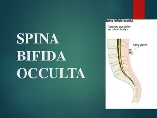
TYPES OF SPINA BIFIDA 1. SPINA BIFIDA OCCULTA--- Failure of fusion of vertebral arches. Skin pigmentation Dimples Sinuses Pedunculated tail Hyertrichosis
TYPES OF SPINA BIFIDA 2. SPINA BIFIDA APERTA Meningocele Meningomyelocele Rachischiais
3. TETHERED CORD SYNDROME. Lipomyelomeningocele Thickened Filum Terminale
4. SPLIT CORD MALFORMATIONS Diastematomyelia Diplomyelia
5. CONGENITAL DERMAL SINUS 6. ANTERIOR MENINGOCELE
SPINA BIFIDA OCCULTA - PATHOGENESIS Failure of fusion of vertebral arches Most frequent in the 5th lumbar and 1st sacral May involve only one vertebra or may extend over several segments.
SPINA BIFIDA APERTA MYELOMENINGOCELE Almost all patients with myelomeningocele also have Chiari II hindbrain malformation Associated medullary kinking, tectal beaking and intrinsic nuclei abnormalities. brainstem defects include
PATHOGENESIS OF MYELOMENINGOCELE Closure of the posterior neuropore occurs at approximately 26 days of gestation. According to the most widely accepted theory, the posterior neuropore fails to close completely during neurulation, and a myelomeningicele, malformation and hydrocephalus develop. Chiari II Toxic agents like cytochalasin, vinblastine, calcium channel antagonists, phospholipase C, concanavalin A, retinoic acid, hydroxyurea and mitomycin C have been implicated in experimental studies carried out in animals.
PATHOGENESIS OF MYELOMENINGOCELE A unified hypothesis to explain the sequential development of myelomeningocele and Chiari II malformation have been proposed. In normal development during the period of rapid brain enlargement after the closure of the posterior neuropore, the neurocele (primitive central canal) occludes transiently. If the posterior neuropore fails to close, causing a myelomeningocele, the neurocele fails to occlude. Without this occlusion, the CSF flows out through the defect in the open posterior neuropore.
PATHOGENESIS OF MYELOMENINGOCELE With the lack of distension of the brain by the CSF, formation of the cranium and its contents are disrupted. The posterior fossa is small, and both upward and downward herniation of the cerebellum occurs, resulting in Chiari II malformation.
MYELOCYSTOCELE Terminal myelocystoceles are skin covered midline masses that occur in the lumbosacral area and are distinct from meningomyeloceles. Myelocystoceles account for 5% of skin covered masses and most likely represent one end of the spectrum of lesions, with lipomyelomeningoceles at the other end. Alternative terms for myelocystocele are syringocele, syringomyelocele and lipomeningomyelocystocele. Myelocystoceles are often associated with hydromyelia and cloacal exstrophy.
MYELOCYSTOCELE Cloacal exstrophy, also called vesicointestinal fissure, is a severe malformation of the abdomen and pelvis with an omphalocele, bladder exstrophy, ambiguous genitalia, imperforate anus and pelvic abnormalities. When all these abnormalities are present, the defect is called OEIS Complex (omphalocele, bladder exstrophy imperforate anus and spinal defect).
PATHOGENESIS OF MYELOCYSTOCELE Myelocystoceles are often associated with cloacal exstrophy. During embryogenesis, the cloaca is an endoderm lined chamber that receives the hindgut and allantois. The cloacal membrane, a bilaminar veil of ectoderm and endoderm, divides the cloaca. Cloacal exstrophy occurs from the rupture of the cloacal membrane and failure of the lateral abdominal wall to close.
PATHOGENESIS OF MYELOCYSTOCELE According to one theory, herniation of the lower abdomen and pelvis disturbs the notochord secretion of inductive factors that regulate the development of neural tube and retrogressive differentiation. Another theory suggests that the CSF is unable to exit from the early neural tube as it should, and it distends the distal terminal ventricle after canalization. The terminal ventricle distends the surrounding dorsal membrane and arachnoid, causing the surrounding meningocele.
PATHOGENESIS OF MYELOCYSTOCELE Teratogens that cause spina bifida aperta in hamsters, such as retinoic acid, also causes lesions resembling myelocystoceles. Most likely the pathogenesis reflects both mechanical disruption of normal development as well as lack of appropriate induction of differentiation factors.
MENINGOCELE Sessile, sometimes pedunculated and are of variable size. Covered by full thickness skin. No evidence of neural tissue or cells. No neurological deficit.
TETHERED CORD SYNDROME EMBRYOLOGY Tip of the conus is below the lower border of L1 vertebral body. It occurs when there is dysfunction before the completion of the neural tube closure, or if the closure is faulty, or the mesenchymal cells enter the central canal of the neural tube.
TETHERED CORD SYNDROME EMBRYOLOGY The mesenchymal cells differentiate into fatty tissue to form lipomyelomeningocele. Marin - Padilla described dural schisis as the basic defect resulting in various forms of tethering, with a short thick filum terminale occurring as a secondary event.
TETHERED CORD SYNDROME PATHOGENESIS The cord is tethered. Nerve roots lie lax and loosely on either side.
TETHERED CORD SYNDROME PATHOGENESIS Effects is maximum close to the site of tethering. When tethering extends over a distance, the effect is maximum on the cord adjacent to the caudal end of the lesion
TETHERED CORD SYNDROME PATHOGENESIS It occurs whenever there is increase in tension in the tethered cord. This occurs by elongation of the spinal canal, in children, during periods of rapid growth, and in adukts during activities or accidents which flex the spine or the hip joints.
SPLIT CORD MALFORMATIONS EMBRYOLOGY Diastematomyelia is a developmental defect of the neuraxis and the spinal column. It is characterised by a division of the spinal cord or cauda equina into two distinct compartments by a septum of bones, cartilage or fibrous tissue.
SPLIT CORD MALFORMATIONS EMBRYOLOGY It is invariably associated with other anomalies of the vertebral body or the neural arches.
SPLIT CORD MALFORMATIONS PATHOGENESIS Two hypotheses were proposed: a) Hydrocehalomyelic hypothesis of Gardner. b) Bremer s hypothesis attributes the defect to a partial obliteration neurenteric canal Pang proposed a unified theory to explain both diastematomyelia and diplomyelia. of an accessory
SPLIT CORD MALFORMATIONS PATHOGENESIS Pang s unified theory states that a persistent endomesenchymal fistula between the amniotic cavity and yolk sac lined by endoderm and an outer condensation of surrounding totipotential mesenchymal cells split the vertebra, notocord and the neural tube.
SPLIT CORD MALFORMATIONS PATHOGENESIS The hemicords may lie within two dural sleeves separated by a rigid osseocartilaginous spur (Type I SPLIT CORD MALFORMATION) A single dural sleeve may enclose the hemicords separated by a fibrous septum (Type II SPLIT CORD MALFORMATION)
ANTERIOR MENINGOCELE Anterior sacral meningocele
ANTERIOR SACRAL MENINGOCELE PATHOGENESIS The meninges protrude into the pelvis causing compression of the pelvic organs May present as a neurogenic bladder. Intrathoracic meningoceles are often associated with neurofibromatosis
CONGENITAL DERMAL SINUS EMBRYOLOGY A developmental defect characterised by a tract lined by squamous epithelium extending from the surface ectoderm terminating at varying depths and occasionally extending to the spinal cord or medulla. Arises as a result of failure of separation of neuroectoderm from the epithelial or surface ectoderm (which normally takes place in the 3rd-5th week of intrauterine life).
CONGENITAL DERMAL SINUS PATHOGENESIS Poses a neurosurgical problem when it extends into the spinal canal / cranial cavity: a) Proves to be a potential source of infection b) Distorts growth of the neural tissues c) Acts as a space occupying lesion when the inner end distends to form an epidermoid or dermoid cyst.
THANK YOU THANK YOU