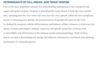
Overview of Corynebacterium diphtheriae Pathogenesis and Virulence Factors
Corynebacterium diphtheriae is a non-spore forming Gram-positive bacillus responsible for causing diphtheria in humans. This bacterium, particularly C. diphtheriae, is known for producing a potent exotoxin called diphtheria toxin, which blocks protein synthesis and leads to serious complications such as demyelinating peripheral neuritis and myocarditis. The pathogenesis involves bacterial adherence, multiplication, and local tissue necrosis, eventually resulting in the formation of a characteristic pseudomembrane. Understanding the mechanisms of action, virulence factors, and cultural characteristics helps in the identification and management of C. diphtheriae infections.

Download Presentation

Please find below an Image/Link to download the presentation.
The content on the website is provided AS IS for your information and personal use only. It may not be sold, licensed, or shared on other websites without obtaining consent from the author. Download presentation by click this link. If you encounter any issues during the download, it is possible that the publisher has removed the file from their server.
E N D
Presentation Transcript
Medical microbiology Lec. 8 Non-spore forming Gram- positive rods By: Lect. Shaima a Al-Salihy
Corynebacterium: - Many members corynebacterium are members of the normal flora of the skin and mucous membranes of the humans. Non-spore forming Gram-positive bacilli of the genus Corynebacterium - Corynebacterium diphtheriae, is the most important member of the group, it can produce a powerful exotoxin that causes diptheria in human. - Nondiphtherial collectively diphtheroids originally were believed to be mainly contaminants. corynebacteria referred to as
Morphology & identification: Corynebacterium - G+ve rods, clupped-shaped (swollen ends), arranged in acute angles (V or L shape, pallisade, Chinese letter pattern). - Have beaded (due to the metachromatic granules). - Non-motile. cuneiform, appearance
Cultural characteristics: - Aerobic and bacteria. - Loaffler s serum slope: the growth is very fast and produce in 6 8 hours, the colonies are small, circular, opaque, and white, at a temperature of 37 C. facultative anaerobic - Tinsdale media: grayish black colonies surrounded by a dark brown halo due to H2S production (Diagnostic).
Cultural characteristics: - MacLeod s (potassium tellurite) blood agar: gray or black colored colonies after 48 hours of incubation. - Mitis colonies are small, round, convex, and black. Intermedius colonies are small, flat, and gray. Gravis colonies are large, irregular, and gray. C. diphtheriae biotypes :
Mechanism of action Virulence factor: Diphtheria toxin: - Neuro- and cardiotoxin - Heat-labile toxin. - Blocks protein synthesis - Encoded introduced bacteria with lysogenic phage Beta-corynephage - Controlled repressor protein. - Expressed if [iron] low. - Composed of fragment A & B B for transport A inhibits protein synthesis. by (Tox lysogenized gene) in by bacterial
Pathogenesis: - Site of entry is respiratory tract but may enter through skin or eye. - Adherence and multiplication of the bacteria in the epithelial cells of infected region, forming a local lesion. - Secrete exotoxins that cause necrosis of the cells in that area. - Formation of the characteristic pseudomembrane. - C. diphtheriae does not cause any invasion of the cell, but the toxin may absorbed into the blood stream and distributed, resulting in diphtheria including demyelinating peripheral neuritis and myocarditis. systemic complications of
C. diphtheria Clinical findings: - The clinical diphtheria depend upon the following: Immune status of the patient. Virulence of the bacteria. The site of the infection. manifestations of Respiratory diphtheria: - The incubation period is 2-5 days. - Sore throat, fever, dyspnea, marked edema of the tonsils, uvula, and anterior neck (bull neck). - formation of pseudomembrane. * Nontoxigenic strains (tox+) cause a mild disease, such as cutaneous diphtheria
Laboratory diagnosis: throat swab Laboratory diagnosis: Specimens: pseudomembrane Microscopy: - Staining by Gram or methylene blue (G +ve rods arranged as L or V shaped) - Albert stain: differential metachromatic granules. Culture: - cultivated on Loeffler s slant, nonselective media (e.g., blood agar) as well as on selective media (e.g., tellurite agar). or swab from stain for
16S probes identification of genus and species of Corynebacteria. Toxigenicity test: 1 1- - in in vivo 2 2- - in in vitro precipitation test to confirm toxin production. 3 3- - tissue 4 4- - PCR isolated from the patient. ribosomal have ribonucleic been acid (rRNA) for designed the vivo: : animal inoculation, Schick test. vitro: : (Elek s test) antibody-based gel diffusion tissue culture PCR assay culture neutralization assay. assay: : for the presence of toxin gene in the organism
Treatment: Treatment should be started immediately after the clinical diagnosis of diphtheria. Treatment of diphtheria is based on: 1. Antitoxin therapy 2. Antibiotics therapy Prevention and Control: Active diphtheria toxoid (combined with tetanus toxoid and acellular pertussis (triple DTaP vaccine) is the key in preventing diphtheria. immunization by vaccination with
Epidemiology: C. diphtheria Either clinical or subclinical infection at an early age yield protective levels of antitoxin. Thus most members of the population, except children are immune. By age 6-8 years about 75% of children in developing countries where skin infection with diphtheria are common have protective serum antitoxin. Absorption of small amount of C. diphtheria toxin does not produce a disease, but it can serve as antigenic stimulus for immune system to produce antitoxin. Active immunization with C. diphtheria toxoid during childhood yield antitoxin levels that generally adequate until adulthood. Young adults in developed countries (Non endemic areas) should receive a booster dose because diphtheria bacilli are not sufficiently prevalent to provide a stimulus of subclinical infection. DPT is the combined of diphtheria toxoid, tetanus toxoid & pertussis vaccine used as a single injection for immunization of children. For booster dose in adults only TD is used.
Corynebacterium jeikeium Opportunistic infections ( sepsis, endocarditis) in immunocompromised (e.g., disorders, bone marrow catheters) Multiple antibiotic resistance common (MDR) Carriage on skin of up to 40% of hospitalized patients (e.g., marrow t-plants) patients transplants, with intravenous blood Corynebacterium urealyticum Urinary tract infections (UTI s); cause chronic or recurrent cystitis, bladder stones, and pyelonephritis rare but important Urease hydrolyzes urea; release of NH4+, increase in pH, alkaline urine, renal stones REVIEW
Listeria monocytogenes Morphology & identification: - short G+ frequently occurs in chains. - Non-spore forming. - Multiply temperatures (4oC) - It has a tumbling end-over-end motility at room temp., but not at 37 C. - It is facultative anaerobe, catalase positive, CAMP positive. Culture & growth : - Muller-Hinton agar. - On blood agar (sheep RBCs) small zone of hemolysis may be observed. Listeria monocytogenes rods (coccobacilli), at refrigerator
Pathogenesis: - route of transmission is eating food cntaminated with the bacteria. - Group at risk are primarily pregnant women, newborns, & adults with weakened immune systems. - L. monocytogenes enters the body through the contaminated food e.g. Cheese or vegetables. - Enters the epithelial cells by induced phagocytosis. and start to produce listeriolysin O. Listeria monocytogenes GIT with the
Listeria monocytogenes Clinical findings: - Intrauterine infection results in sepsis and death before or after delivery. - Meningitis: between the birth and the 3rd. week of life. - Meningitis is often complicated by encephalitis, a pathology that is unusual for bacterial infections.
Intracellular Survival & Replication of Listeria Phagocytosis Macrophage Listeriolysin O? Macrophage Intracellular Replication Actin Filaments REVIEW
Epidemiology of Listeria Infections Natural Reservoirs Common Routes for Human Exposure Population at Greatest Risk REVIEW
Actinomycetes Are a diverse group of G+bacilli with a tendency to form chains or filaments. Most are saprophytes that lives in soil, others are normal flora. Actinomyces israelii: - Non-acid fast, Branching rods. - Normal flora of gingival crevices and female genital tract Pathogenesis: - Causes actinomycosis: granulomatous, generally invasive, penetrating all tissues, including bone. - Tissue swelling draining abscesses (sinus tracts) with "sulfur" granules(hard yellow microcolonies) in exudate that can be used for microscopy or culture and composed of macrophages, tissue cells, fibrin and bacteria. Actinomyces israelii chronic not suppurative painful & but very
laboratory diagnosis: - Specimen: Pus from draining sinuses, sputum, or specimens from tissue. - Microscopy: examined for the presence of sulfur granules (Diagnostic). - culture: Brain heart infusion agar and thioglycolate broth and incubated anaerobically or elevated CO2 conditions.
Nocardia: - Nocardia asteroides, Nocardia brasiliensis Morphology: - Aerobic - Gram-positive branching rods, partially acid fast. Pathogenesis - No toxins or virulence factors known - an opportunistic infection associated with several risk factors, most of which impair the cell-mediated immunity. - Pulmonary nocardiosis may disseminate to other organs (brain or skin). - The usual pathologic process (neutrophilic inflammation). - Cutaneous/subcutaneous nocardiosis: swelling draining subcutaneous abscesses with granules (mycetoma) is abscess formation cellulitis with
laboratory diagnosis: - Culture of Clinical specimen :Sputum, Pus, Biopsy tissue
Case study: An 8-year-old child was admitted to hospital with low-grade fever (38 C), malaise, dysphagia, and headache. Examination revealed a fibrinous membrane on the pharynx. A pair of swab collected from the membrane was sent to microbiology laboratory for diagnosis of Corynebacterium diphtheriae. The child was administered antitoxin by the intravenous route and treated with antibiotics. Subsequently, 3 days later the report from the laboratory confirmed isolation of a toxigenic C. diphtheriae from the specimen. Why treatment for diphtheria was started immediately without waiting for the confirmatory report? What are the tests performed in a microbiology laboratory to demonstrate C. diphtheriae in clinical specimens? Are nontoxigenic strains of C. diphtheriae capable of causing disease in humans?