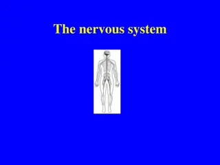
Understanding the Physiology of the Spinal Cord
The spinal cord serves as the information highway between the brain and body, allowing for sensory input reception and motor signal transmission. It plays a crucial role in locomotion, reflex responses, and overall coordination of muscle activities. The cylinder of nerve tissue within the vertebral canal extends to L1 in adults, giving rise to 31 pairs of spinal nerves. The fibrous layers enclosing the spinal cord provide protection and support, with gray matter containing neuron cell bodies and white matter consisting of myelinated axons.

Download Presentation

Please find below an Image/Link to download the presentation.
The content on the website is provided AS IS for your information and personal use only. It may not be sold, licensed, or shared on other websites without obtaining consent from the author. Download presentation by click this link. If you encounter any issues during the download, it is possible that the publisher has removed the file from their server.
E N D
Presentation Transcript
Physiology of Spinal Cord Compiled By Compiled By Prof. Sudhir Kumar Awasthi Dept. Of Life Sciences Prof. Sudhir Kumar Awasthi Dept. Of Life Sciences CSJMU CSJMU
Information highway between brain and body Extends through vertebral canal from foramen magnum to L1 Each pair of spinal nerves receives sensory information and issues motor signals to muscles and glands Spinal cord is a component of the Central Nervous System while the spinal nerves are part of the Peripheral Nervous System
Conduction bundles of fibers passing information up and down spinal cord Locomotion repetitive, coordinated actions of several muscle groups central pattern generators are pools of neurons providing control of flexors and extensors (walking) Reflexes involuntary, stereotyped responses to stimuli (remove hand from hot stove) involves brain, spinal cord and peripheral nerves
Cylinder of nerve tissue within the vertebral canal (thick as a finger) vertebral column grows faster so in an adult the spinal cord only extends to L1 31 pairs of spinal nerves arise from cervical, thoracic, lumbar and sacral regions of the cord each cord segment gives rise to a pair of spinal nerves Cervical and lumbar enlargements Medullary cone (conus medullaris) = tapered tip of cord Cauda equinae is L2 to S5 nerve roots resemble horse s tail
3 Fibrous layers enclosing spinal cord Dura mater tough collagenous membrane surrounded by epidural space filled with fat and blood vessels epidural anesthesia utilized during childbirth Arachnoid mater layer of simple squamous epithelium lining dura mater and loose mesh of fibers filled with CSF (creates subarachnoid space) Pia mater delicate membrane adherent to spinal cord filium terminale and denticulate ligaments anchor the cord
Central area of gray matter shaped like a butterfly and surrounded by white matter in 3 columns Gray matter = neuron cell bodies with little myelin White matter = myelinated axons
Pair of dorsal or posterior horns dorsal root of spinal nerve is totally sensory fibers Pair of ventral or anterior horns ventral root of spinal nerve is totally motor fibers Connected by gray commissure punctured by a central canal continuous above with 4th ventricle
White column = bundles of myelinated axons that carry signals up and down to and from brainstem 3 pairs of columns or funiculi dorsal, lateral, and anterior columns Each column is filled with named tracts or fasciculi (fibers with a similar origin, destination and function)
Ascending and descending tract head up or down while decussation means that the fibers cross sides Contralateral means origin and destination are on opposite sides while ipsilateral means on same side
Pain signals from tissue injury Decussate in spinal cord and ascend with spinothalamic fibers End in reticular formation (medulla and pons) 3rd and 4th order neurons continue to thalamus and cerebral cortex 13- 10
Proprioceptive signals from limbs and trunk travel up to the cerebellum Second order nerves ascend in ipsilateral lateral column
31 pairs of spinal nerves (1st cervical above C1) mixed nerves exiting at intervertebral foramen Proximal branches dorsal root is sensory input to spinal cord ventral root is motor output of spinal cord cauda equina is roots from L2 to C0 of the cord Distal branches dorsal ramus supplies dorsal body muscle and skin ventral ramus to ventral skin and muscles and limbs meningeal branch to meninges, vertebrae and ligaments Spinal nerves: 8 cervical, 12 thoracic, 5 lumbar, 5 sacral and 1 coccygeal. Each has dorsal and ventral ramus.
Ventral rami branch and anastomose repeatedly to form 5 nerve plexuses cervical in the neck, C1 to C5 supplies neck and phrenic nerve to the diaphragm brachial in the armpit, C5 to T1 supplies upper limb and some of shoulder and neck lumbar in the low back, L1 to L4 supplies abdominal wall, anterior thigh and genitalia sacral in the pelvis, L4, L5 and S1 to S4 supplies remainder of lower trunk and lower limb coccygeal, S4, S5 and C0
Quick, involuntary, stereotyped reactions of glands or muscle to sensory stimulation automatic responses to sensory input that occur without our intent or often even our awareness Functions by means of a somatic reflex arc stimulation of somatic receptors afferent fibers carry signal to dorsal horn of spinal cord one or more interneurons integrate the information efferent fibers carry impulses to skeletal muscles skeletal muscles respond
Sense organ (proprioceptor) that monitors length of muscle and how fast muscles change in length Composed of intrafusal muscle fibers, afferent fibers and gamma motorneurons
When a muscle is stretched, it contracts and maintains increased tonus (stretch reflex) helps maintain equilibrium and posture head starts to tip forward as you fall asleep muscles contract to raise the head stabilize joints by balancing tension in extensors and flexors smoothing muscle actions Very sudden muscle stretch causes tendon reflex knee-jerk (patellar) reflex is monosynaptic reflex testing somatic reflexes helps diagnose many diseases Reciprocal inhibition prevents muscles from working against each other
10-12,000 people/ year are paralyzed 55% occur in traffic accidents This damage poses risk of respiratory failure Early symptoms are called spinal shock Tissue damage at time of injury is followed by post-traumatic infarction
Neuroanatomy & Neurophysiology: a review by J.S. Citow, R.L MacDonald (2004) The Human Nervous system:An anatomical viewpoint by Murray Barr(2000) Textbook of Clinical Neuroanatomy by Vishram Singh (2007)


")


 that monitors length of")