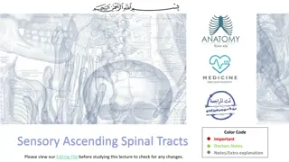
Spinal injuries

An overview of spinal injuries, including anatomy revision, types of spinal fractures, assessment, management, and common patterns of injuries. It also covers back pain presentation, red flags, differentials, and the structure of vertebrae. The Denis classification of spinal fractures and the most common types of spinal fractures are explored.

Download Presentation
Please find below an Image/Link to download the presentation.
The content on the website is provided AS IS for your information and personal use only. It may not be sold, licensed, or shared on other websites without obtaining consent from the author. Download presentation by click this link. If you encounter any issues during the download, it is possible that the publisher has removed the file from their server.
Presentation Transcript
Spinal injuries Katherine Flack 4thyear medical student Tim Gardner Orthopaedic Registrar
For IT Text in blue indicates a question for students to answer Information regarding how the question will work (eg free text of multiple choice) is listed in the notes section of the slide References are also in the notes section Slides titled navigation is how the navigation pane should appear on the side
Navigation Anatomy revision Overview of spinal fractures - Denis classification - Common types Compression fracture case - Situation and background - Assessment - Overview of compression fractures Traumatic spinal cord injury (TSCI) case - Situation and background - C-spine immobilisation - Assessment - Management 1 - Management 2 - Summary of TSCI - ASIA chart - Patterns of injuries - Management 3
Navigation part 2 Back pain - Presentation - Red flags - Differentials Summary
Spinal column anatomy 33 bones separated by intervertebral discs
Structure of vertebrae Anterior ventral body weight bearing Posterior vertebral arch multiple bony prominences which enable the attachments for muscles and ligaments
Denis classification of spinal fractures The Denis Classification of spinal fractures divides the spine into 3 columns the anterior, middle and posterior Anterior column anterior longitudinal ligament and anterior half of vertebral body Middle column posterior half of vertebral body and posterior longitudinal ligament Posterior column pedicles, facet joints and supraspinous ligaments If 2 or more of these columns are injured the spine is described as unstable
Most common types of spinal fractures Click on the boxes to find out more about type of spine of spinal fracture Chance Burst Compression (flexion/distraction) Anterior part of vertebra breaks but the posterior aspect remains intact therefore only loses height anteriorly. Vertebra loses height on both anterior and posterior aspects. Occurs when vertebrae are suddenly pulled away from each other in a flexion-distraction mechanism. Usual cause is falling from height and landing on feet. Associated with high rates of mechanical instabilities and gastrointestinal injuries. Commonly occurs in patients with osteoporosis.
Situation and background Mary is a 75 year old lady who lives alone in a bungalow. She has a PMHx of acid reflux, osteoporosis, type 2 diabetes. Whilst out gardening she slipped on the steps outside. What are you worried about when an elderly patient presents with a fall? Fracture Head injury Collapse due to underlying cause eg stroke, MI, hypoglycaemia, rupture abdominal aortic anerusym
Assessment When she arrives in A&E she is sent for an urgent x-ray which confirms your suspicions of a compression fracture. Select which aspect of her past medical history is most likely to have contributed to her injury? - Acid reflux - Female - Osteoporosis - Type 2 diabetes - Age
Compression fracture Compression fractures are common in individuals with osteoporosis. Diagnosis is done using lateral x- rays. On x-ray there is a loss of anterior, middle or posterior vertebral height by 20%. This shows the typical wedge shape. Treatment observations and pain management mostly
Situation and background You are an FY1 working in A&E. Greg (age 35) has arrived to ARI by helicopter after being involved in a serious high speed head-on collision whilst driving. He has been initially managed by trauma doctors at the scene and has a cervical collar and blocks and board in place. He is able to talk and his main complaint to you is pain in neck and tingling in arms.
C-spine immobilsation This is how Greg arrives to you What is the main indication in this case to use a c-spine immobilisation collar and blocks? Paresthesia in extremities Car collision mechanism
Assessment for cervical spine injury NICE recommend using the indications given in the Canadian C-spine rule to determine whether to maintain full in-line immobilisation. Using the Canadian-C spine rule Greg is deemed to be high risk (due to dangerous mechanism). Therefore you should maintain full in-line spinal immobilisation. This is also used to determine if radiography should be used, in this case the answer is yes.
How do you initially manage the patient? Take a thorough history Give pain medication <C>ABCDE approach CT scan
<C>ABCDE Catastrophic bleeding / Cervical Spine Pelvic binder in place, abdomen hard. Neck Immobilisation C Airway There is no airway obstruction, airway patent A Breathing O2 sats at 92 and clear lung sounds. Equal chest expansion, no flail chest B Circulation Blood pressure is 95/60, weak pulse in peripheries. HR 110 bpm, cool peripherally C Disability Pupil responses equal and reactive, numbness and weakness in right arm D Exposure/everything else Glucose levels normal, multiple grazes and abdominal bruising but no other major external injuries E
Next management Thinking about each component of the <C>ABCDE approach, list how you would now like to manage Greg? Circulation IV fluid resuscitation start with IV Crystalloid but consider early switch to blood When answering a complex question like this it can be helpful to group into the C ABCDE order. Disability Covered by CT scan Neurological exam Catastrophic bleeding IV access Bloods group and save (pink tube), FBC, U+E, Coag Urgent CT scan request camp bastion protocol Head, neck, chest, abdomen, pelvis at least Everything else Pain relief likely IV Opioid Involve seniors and other specialities Should have a trauma call but just in case, make sure everyone is here! (orthopaedics, general surgery, anaesthetics, radiology) Update family Airways + breathing Oxygen 15L high flow non-rebreathe mask
Images to be included on page when answers appear from the previous slide
CT scan After the initial handover and <C>ABCDE, Greg is sent for a full CT scan. Findings C5/6 Facet joint dislocation Free fluid in abdominal cavity, suspicious of splenic rupture No pelvic fracture No pneumothorax, no rib fractures No intracerebral injuries No other bony injuries
Traumatic spinal cord injury (TSCI) More common in males. Most are due to preventable causes such as RTA, sport injuries or falls. Can be classified as complete or incomplete (AOSpine Injury Classification System). Complete damage affects the whole spinal cord width which causes complete loss of sensation and paralysis below the level of injury. Incomplete damage only affects part of the spinal cord, hence only partially implicating sensation or movement below the level of injury.
Pathophysiology of TSCI Primary injury the direct trauma from injury mechanism causes damage to spinal cord. Secondary injury result of injuries to surrounding structures causing compression on the spinal cord often from haematoma.
Spinal surgeon The spinal orthopaedic surgeon is sent to review Greg. First she goes through the ASIA chart to determine the level of severity of neurological deficit.
ASIA chart Developed as a universal classification tool for spinal cord injuries. Grades A: complete no sensory or motor function preserved B: sensory incomplete sensory but not motor function is preserved C: motor incomplete less than half of key muscle functions below the lesion have a muscle grade of >/= 3 D: motor incomplete half or more key muscles functions below the lesion having a muscle grade >/= 3 E: normal For more in depth classification look up the ASIA Impairment Scale (AIS).
Gregs neurological injuries Greg has these findings on neurological examination especially: Anal tone normal Weakness to wrist extension bilaterally Numbness and tingling in thumb bilaterally At which spinal level do these injuries correspond to? - C2/3 - C3/4 - C5/6 - C6/7
ASIA Impairment Scale (AIS) Based off of Greg s injuries and ASIA chart, what AIS grade do you give him? A B C D E He has an incomplete TSCI as the damage only affects part of the spinal cord, hence only partially implicating sensation or movement below the level of injury. He is not paralysed below the level of injury indicating it is not a complete TSCI.
Patterns of injuries Tetraplegia impairment of function in arms, trunk, legs and pelvic organs. Paraplegia impairment of function in trunk, legs and pelvic organs. Arm function is preserved.
Management Spinal column injuries can be managed non-operative or operative. There are 2 absolute indications for surgical management of TSCI - Progressive neurological deficit Dislocation type injury to spinal column What will the spinal surgeon s decision be regarding Greg s management? - Operate
Rehab After stabilization of his major bleeding, he then has successful spinal surgery and Greg is now in rehab. Which members of the multi-disciplinary team may be involved in his care? Medical team Nurses Physiotherapy Speech and language Occupational therapy Psychologist
Back pain presentation John is a 55 year old man who presents to his GP with back pain.
Using a systematic approach, which questions would you initially like to ask regarding the pain? Using SOCRATES Site Onset Character Radiating pain Associated symptoms Time/duration Excerbating/relieving factors Severity
Back pain presentation John replies with these answers - Pain is in lower back It came on approximately a week ago after lifting a heavy box in work Pain is dull and does not radiate Regular ibuprofen helps a bit Rates the pain an 7/10 and was at it s worst the day after lifting the box
Back pain red flags Which red flags questions should you ask to rule out a more serious cause of the back pain? Cauda equina red flags - Bilateral sciatica - Bilateral motor weakness of legs - Difficulty initiating micturition - Faecal incontinence - Saddle anesthesia - Decreased anal tone - Erectile dysfunction Other red flags - Night pain - Stiffness in morning - Major trauma - Gradual onset - Weight loss - Fever - History of cancer
Differentials of back pain There are many differentials of back pain, try list as many as you can
Differentials of back pain Mechanical - Muscle of ligament sprain - Herniated disc - Scoliosis - Degenerative changes Spinal fracture Cancer Spinal infection Back pain Non-spinal related Eg Ruptured aortic aneurysms, pyelonephritis + more Ankylosing spondylitis Cauda equina Spinal stenosis
John says he is not affected by any of those symptoms. He has a normal PR exam, and no altered perineal sensation. He went to pass urine before coming into the clinic room. You are confident this is likely a mechanical cause and prescribe simple analgesia to John with a worsening statement given.
Summary Be aware of risk factors for compression fractures. Recognise the signs of spinal cord injury and gain confidence using the ASIA chart. Recognise red flag symptoms in back pain which may require urgent investigation.


")


")