Helicobacter Pylori: Characteristics, Pathogenesis, and Diagnosis
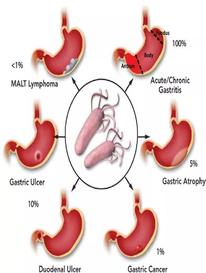
Helicobacter pylori is a spiral-shaped Gram-negative bacterium associated with various gastrointestinal conditions like gastritis, duodenal ulcers, gastric ulcers, and gastric carcinoma. It exhibits unique characteristics in common with Campylobacters and has specific culture and growth requirements. The pathogenesis involves the production of urease activity, enabling survival in the acidic gastric environment. Clinical findings, lab diagnosis methods, and epidemiology of H. pylori infection are also discussed in the context of gastrointestinal illnesses.
Download Presentation

Please find below an Image/Link to download the presentation.
The content on the website is provided AS IS for your information and personal use only. It may not be sold, licensed, or shared on other websites without obtaining consent from the author. Download presentation by click this link. If you encounter any issues during the download, it is possible that the publisher has removed the file from their server.
E N D
Presentation Transcript
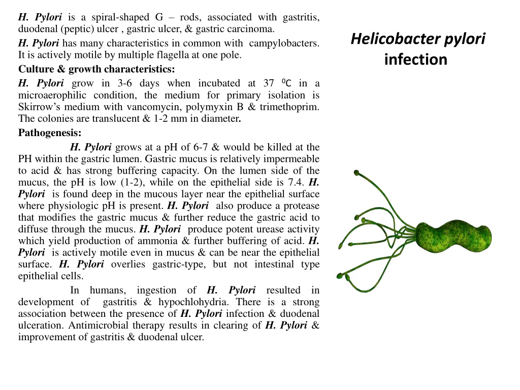
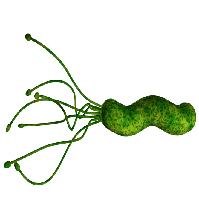
H. Pylori is a spiral-shaped G rods, associated with gastritis, duodenal (peptic) ulcer , gastric ulcer, & gastric carcinoma. H. Pylori has many characteristics in common with campylobacters. It is actively motile by multiple flagella at one pole. Culture & growth characteristics: H. Pylori grow in 3-6 days when incubated at 37 microaerophilic condition, the medium for primary isolation is Skirrow s medium with vancomycin, polymyxin B & trimethoprim. The colonies are translucent & 1-2 mm in diameter. Pathogenesis: H. Pylori grows at a pH of 6-7 & would be killed at the PH within the gastric lumen. Gastric mucus is relatively impermeable to acid & has strong buffering capacity. On the lumen side of the mucus, the pH is low (1-2), while on the epithelial side is 7.4. H. Pylori is found deep in the mucous layer near the epithelial surface where physiologic pH is present. H. Pylori also produce a protease that modifies the gastric mucus & further reduce the gastric acid to diffuse through the mucus. H. Pylori produce potent urease activity which yield production of ammonia & further buffering of acid. H. Pylori is actively motile even in mucus & can be near the epithelial surface. H. Pylori overlies gastric-type, but not intestinal type epithelial cells. In humans, ingestion development of gastritis & hypochlohydria. There is a strong association between the presence of H. Pylori infection & duodenal ulceration. Antimicrobial therapy results in clearing of H. Pylori & improvement of gastritis & duodenal ulcer. Helicobacter pylori infection 0C in a H. Pylori of resulted in
The mechanism by which H. pylori causes mucosal inflammation and damage involve both bacterial & host factors. The bacteria invade the epithelial cells to a limited degree. Toxins & LPS may damage the mucosal cells, & the ammonia produced by the urease activity may directly damage the cells also. Clinical findings: Acute infection gastrointestinal illness with nausea & pain. Vomiting & fever may be present also. The acute symptoms may lasts for less than 1 week or 2 weeks. Once colonized, H. pylori infection persist for years or even lifetime. About 90% of patients with duodenal ulcer & 50-80% of those with gastric ulcer have H. pylori infection. H. pylori may have a role in gastric carcinoma & lymphoma. Lab. Diagnosis: Specimens: gastric biopsy can be used for histological examination or can be minced in saline & used for culture. Serum for demonstration ofAbs. Smear: The diagnosis of gastritis & H. pylori infection can be made histologically. A gastroscopy procedure with biopsy is required. Routine stain demonstrate gastritis & Giemsa or special silver stains can show the curved or spiral bacteria. Helicobacter pylori infection can yield an upper
Culture: on selective media & incubation conditions. Serology: serum Abs specific for H. pylori infection may persist even if the infection is eradicated, so serology is of limited value in the diagnosis. Other tests: Tests to detect urease activity are widely used for presumptive identification of H. pylori infection in gastric biopsy. Detection of H. pylori Ag in stool is used to monitor treated patients with known infection by H. pylori. Immunity: Patients infected by H. pylori develop IgM Ab response, followed by IgG & IgA , & these persist both systemically & at the mucosa in high titers in chronically infected patients. Epidemiology: H. Pylori is present on the gastric mucosa of less than 20% of persons under age 30, & increases to 40-60% in those age 60 years. Including asymptomatic. In developing countries, the prevalence of infection may be 80% or higher in adults. Person to person transmission of H. pylori is likely because intrafamilial clustering of infection occurs. Helicobacter pylori infection those who are
