Embryology of the GI Tract and Development of the Gut Tube
Embryology of the gastrointestinal (GI) tract involves the formation of germ layers during gastrulation, leading to the development of endoderm, mesoderm, and ectoderm. By incorporating the yolk sac, the primitive gut tube emerges during weeks 3-4 and is divided into foregut, midgut, and hindgut, giving rise to various digestive structures. Histologically, the adult GI tract consists of different layers derived from endoderm and mesoderm. Abnormalities during development can lead to issues like stenosis and atresia. Specific organs such as the stomach, liver, gallbladder, and pancreas have distinct developmental pathways.

Download Presentation

Please find below an Image/Link to download the presentation.
The content on the website is provided AS IS for your information and personal use only. It may not be sold, licensed, or shared on other websites without obtaining consent from the author. Download presentation by click this link. If you encounter any issues during the download, it is possible that the publisher has removed the file from their server.
E N D
Presentation Transcript
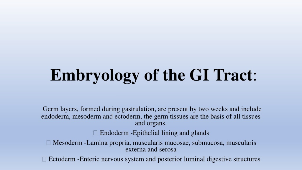
Embryology of the GI Tract: Germ layers, formed during gastrulation, are present by two weeks and include endoderm, mesoderm and ectoderm, the germ tissues are the basis of all tissues and organs. Endoderm -Epithelial lining and glands Mesoderm -Lamina propria, muscularis mucosae, submucosa, muscularis externa and serosa Ectoderm -Enteric nervous system and posterior luminal digestive structures
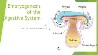
The Primitive gut tube develops during week 3-4 by incorporating the yolk sac during craniocaudal and lateral folding of the embryo. The tube is divided into 3 distinct sections; foregut, midgut and hindgut. Foregut gives rise to the esophagus, stomach, liver, gallbladder, bile ducts, pancreas and proximal duodenum. The midgut develops into the distal duodenum, jejunum, ileum, cecum, appendix, ascending colon, and proximal 2/3 of transverse colon. The hindgut becomes the distal 1/3 of the transverse colon, descending colon, sigmoid colon and the upper anal canal.
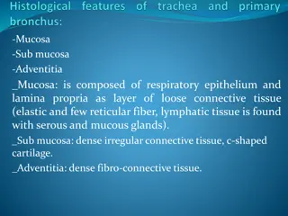
Histologically, The general plan of the adult gastrointestinal tract consists of a mucosa (epithelial lining and glands, lamina propria, and muscularis mucosae), submucosa, muscularis externa, and adventitia or serosa. Embryologically, the epithelial lining and glands of the mucosa are derived from endoderm, whereas the other components are derived from visceral mesoderm. Early in development, the epithelial lining the gut tube proliferates rapidly and obliterates the lumen. Later, recanalization occurs
Formatted: CenteredImage from http://www.med.umich.edu/ Proliferation of the epithelial lining of the gut tube results in obliteration of the lumen by week 6. The central cells then degenerate and the tube is recanalized by week 8.Abnormalities in this process result in: stenosis, atresia, and duplications. Foregut FormationThe foregut gives rise to the esophagus, stomach, liver, gallbladder, pancreas and the caudal portion of the duodenum. Lateral grooves invaginate during week 4 on each side of the proximal foregut and fuse creating the tracheoesophageal septum. The septum separates the respiratory and digestive tracts with the ventral portion developing into respiratory system and dorsal into gastrointestinal tract. By week 16 the esophagus has stratified squamous epithelium and swallow can appreciated.Failure of the tracheoesophageal septum developmentresults in tracheoesophageal fistula and/or esophageal atresia. The stomach develops from a fusiform dilation in the foregut during week 4.
The stomach develops from a fusiform dilation in the foregut during week 4. A 90 degree clockwise rotation createsthe lesser peritoneal sac. The liver develops from an endodermal outgrowth, hepatic diverticulum, at the cranioventral portion of the foregut.Mesoderm surrounds the diverticulum, septum tansversum. Hepatic cells (hepatoblasts), both hematopoietic and endothelial precursor cells, then migrate into the septum tansversum. The endothelial precursor cells, vitelline veins, are surrounded by hepatic cells forming the hepatic sinusoids. Bi-potential hepatoblasts give rise to both cholangiocytes and hepatocytes.The hepatoblasts in mesenchyme closest to the portal vein form a bi- layered structure, the ductal plate.These cells remodel to form bile ducts in the intrahepatic portal tracts. Abnormal development of intrahepatic bile ducts due to ductal plate malformations are likely the underlying cause of congenital hepatic fibrosis and cystic kidney diseaseas well as ciliopathies such asJoubert syndrome, Meckel- Gruber and Ivemark syndrome.
Gallbladder and bile ducts begin as a cystic diverticulum. The gallbladder is initially solid and become cystic.Intrahepatic bile duct development starts at the hilum and progresses tothe periphery of the liver.The common bile duct forms in an area of narrowing between the foregut and the hepatic diverticulum. At birth the most peripheral intrahepatic bile ducts are immature with persistence of ductal plate.Maturity of intrahepaticbiliary tree is achieved by 4 weeks of life.
Pancreas development begins during the 4th-5th weeks of gestation as distinct dorsal and ventral buds arising from the endoderm of the caudal foregut, the proximal duodenum. The dorsal bud is larger than and slightly more cranial to the ventral bud. Each bud communicates with the foregut through a duct. Rotation of the duodenum causes the ventral pancreatic bud to rotate clockwise to the left of the duodenum and brings it posterior and inferior to the dorsal pancreatic bud. The two buds fuse to form the pancreasduring the 7thweek of gestation..
The ventral bud forms the inferior part of the head of the pancreas and the dorsal bud forms the superior part of the head, the body, and the tail of the pancreas. The ductal systems of the two buds fuse in the 8thweek. The main pancreatic duct (duct of Wirsung) which enters the duodenum at the major duodenal papilla (ampulla of Vater)is formed by the longer dorsal duct draining into the proximal ventral duct to form
. If the proximal portion of the dorsal duct remains, it forms an accessory duct (duct of Santorini) that opens into a minor accessory papilla located about 2 cm above the main duct. The accessory duct opens into a minor papilla in 33% of people and ends blindly in 8% of people. Fifty percent of people do not have an accessory duct. Endocrine cells (islets) are identifiable by the 8thweek.Exocrine pancreatic development continues after birth with maturation of specific digestive enzymes
Abnormal development of the pancreas results in several congenital anomalies to include pancreas divisum. This is the most common variant (10%) and results from non-fusion of dorsal andventral ductsduring the second month ofgestation. Annular pancreas is another congenital anomaly. A band of pancreatic tissue encircles the duodenumand is typically associated with other anomalies to includeDown s syndrome and duodenal atresia.
Midgut Formation The distal duodenum, jejunum, ileum, cecum, appendix, ascending colon, and proximal 2/3 of transverse colon develop from the midgut, between the 6 and 10thweeks. The midgut loop herniates through the primitive umbilical ring during umbilical herniation at week 6. By ten weeks of development the abdomen has enlarged so that the entire length of the midgutcan be accommodated. Following a 270 degree counterclockwise rotation around the
superior mesenteric artery, the bowel returns to the abdominal cavity. The large intestine returns following the small intestine and does an additional 180 degree counterclockwise rotation.Colonic fixation occurs afterthe return to the abdomen.Cecum and appendix begin as a diverticulum around the 6thweek.Unequal cecal growth leaves appendix medial to the cecum.
Hindgut Formation (Slides 23-25) The distal 1/3 of the transverse colon, descending colon, sigmoidcolon develop from the cranial end of the hindgut.The upper anal canal also develops from the terminal end of the hindgut with the urorectalseptum dividing the upper anal canal and the urogenital sinus during the 6thweek.
By the 7thweek, theurorectalseptum fuses with the cloacalmembrane, giving rise to the anal membrane and the urogenital membrane. The anal membrane ruptures during the 8thweek allowing communication between the anal canal and the amniotic fluid. The superior 2/3 of the anal canaloriginates from hindgut and the inferior 1/3 is derived from proctodeum.The pectinate line is the junction of proctodeum ectoderm and hindgut endoderm.
The mesentery develops from the mesoderm and connectsthe primitive gut to the body wall. The ventral mesentery is present only between the liver and the stomach, and the liver and the duodenum. It forms the lesser omentum, between the liver and the stomach and duodenum, and the falciformligamentbetween the liver and the anterior body wall. The dorsal mesentery surrounds the rest of the primitive gut. It forms several organ ligaments and also becomes the greater omentum. Finally, the mesentery of the colon develops into the transverse mesocolon. During development some structures come to lie close to the posterior body wall and as the mesentery is absorbedthe organ takes on a retroperitoneal position. Retroperitoneal organs include portionof the duodenum, the pancreas, the ascending and the descending colon.
Blood Supply Appropriate blood supply to the gastrointestinal tract and enteric organs is vital to health. Proximal Esophagus - Inferior Thyroid Artery Thoracic Esophagus - Terminal bronchial arteries Distal Esophagus - Left gastric and left phrenic arteries Stomach -Celiac artery Small intestine- Superior mesenteric artery Large intestine- Superior and Inferiormesenteric arteries
Stomach (Slides 44-49) Gastric Features The stomach serves as a reservoir, mixes and emulsifies food, secretes acids and digestive enzymesand regulates the release of gastric chyme into the duodenum. The fundus acts a reservoir for food. The body is a mixing chamber. The muscular antrum releases small volumes intermittently into the duodenum. Total gastric volume ranges from 30 ml in newborn to 2 L in adults. Gastric Structure The stomach muscle layers includean outer longitudinal layer, a middle circular layer, and an inner oblique layer. The inner lining consists of four layers: the serosa, the muscularis, the submucosa, and the mucosa. Gastric glands are densely packed in the mucosa. The glands contain cells that produce digestive enzymes, hydrochloric acid, and mucus. Gastric cells and their specific func
Gastric pits cellular make-up varies by region Cardiacontains shallow pits with many mucous cells. Few parietal and chief cells. Funduscontains deep, branched pits with mucous cells at the apex. Parietal cells located in the body. Chief and neuroendocrine cells are at the base. Chief cells are only present in the fundus. Antrum contains deep pits with mucus, parietal and neuroendocrine cells.


")


")
")