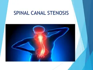
Spinal Immobilization in Trauma: Case Study and Considerations
A 70-year-old male with trauma after falling 20 feet was managed at the ED, demonstrating tenderness in the mid thoracic spine and other injuries. Despite negative cervical spine imaging, the patient underwent interventions for spinal fractures. The decision to transfer without a backboard highlights evolving practices in spinal immobilization. The case underscores the complexities in assessing the need for long backboards and the importance of weighing risks and benefits in spinal trauma management.

Download Presentation

Please find below an Image/Link to download the presentation.
The content on the website is provided AS IS for your information and personal use only. It may not be sold, licensed, or shared on other websites without obtaining consent from the author. Download presentation by click this link. If you encounter any issues during the download, it is possible that the publisher has removed the file from their server.
E N D
Presentation Transcript
70 yr old ambulatory male brought to ED by private vehicle after falling 20 ft from a platform in garage injuring head and chest. Brief LOC, has mild back pain, no neck pain. Level 2 Trauma team activated in triage Exam: BP 128/105, HR 140, RR 28, SpO2 93% GCS 15
Exam: Tenderness in the mid thoracic spine and right para-scapular region. Neck - non tender with full ROM Neuro exam normal FAST negative Chest xray: non displaced right clavicle fx CT head negative CT chest/abd/pelvis with reconstruction T10 vertebral body involving post elements (Chance Fx), hemothorax T8 and T9 spinous processes Right scapular fracture
Transferred to Level 1 trauma center PI Filter: transferred without backboard
CT cervical spine negative Kept on bedrest, TLSO ordered 2ndHD Open reduction of T9-10 fracture with T8-9, T9-10, T10-11, T11-12 Posterior lateral fusion Discharged on 10thhospital day
CT cervical spine? Should this patient have been transferred on a backboard?
Background The benefit of long backboards is largely unproven Long backboards can induce pain, agitation, respiratory compromise, decrease tissue perfusion leading to development of pressure ulcers Use of long backboards should be judicious so benefits outweigh risks
Blunt trauma with altered LOC Spinal pain or tenderness Neurologic complaint (numbness or weakness) Anatomic deformity of the spine High energy mechanism with any of the following: Drug or alcohol intoxication Inability to communicate Distracting injury
Patients with all of the following: Normal LOC (GCS 15) No spine tenderness or anatomic deformity No neurologic findings No distracting injuries No intoxication Penetrating trauma to head, neck or torso with no evidence of spinal injury
Patients who are ambulatory at the scene Patient who must be transported for a protracted time, particularly prior to interfacility transfer
Application of cervical collar Adequate security to a stretcher Minimal movement with transfers Maintenance of in-line stabilization during any necessary movement or transfers
67 yo gentleman group home resident slipped & fell backwards down a flight of stairs. Evaluated in the ED, admitted to the general surgical/trauma unit on Trauma Surgery service then transferred to Neurosurgery the following day (remained on same unit). Found to have spinous process fractures of C7, T1, right facet fracture T1, vertebral body and spinous process fracture T11 and pre-vertebral hematoma in lower thoracic spine. These were managed with a TLSO brace with SOMI extension (ultimately required spinal fusion). 13
He was cleared for upright & out of bed activity while wearing TLSO with SOMI the day after admission HD #2, pt developed respiratory difficulties, was transferred to the SICU and required intubation shortly after. Neurosurgery progress notes documented in PLAN: TLSO with SOMI at all times for next 3 weeks. This was changed in their notes to TLSO w/ SOMI when > 30 degrees or OOB. When < 30 degrees may remove TLSO but will need to use Miami-J . 14
On HD #4, tissue injury noted during skin assessment after removing TLSO with SOMI. Nurse documented Suspected deep tissue injury noted during skin assessment after removing C collar/TLSO brace. Wounds measure 2cm x 1cm and 3cm x 2cm. Foam dressing applied. MD notified. 15
Consult with Certified Wound Ostomy Nurse (CWON) obtained the next morning and they continued to follow/assist with plan of care. Both wounds worsened over the next 3 wks despite efforts to prevent further breakdown. Plastic surgery consult obtained & noted stage of pressure wounds as stage 3 (rt mandible) & stage 4 (lt mandible). In retrospective review of EHR, unable to find order that corresponded with progress note plans & unable to find care plan orders that indicate the device could be removed for routine skin checks & cares per protocol Difficult to find consistent EHR nursing documentation of removal of device for skin assessment & cares. 16
Identifiers: 66 y. o. female fall from standing on gero-psych unit. Filter: Long LOS, readmission, complication PMH: Schizophrenia
66 y. o. year old female who initially presented to the hospital for evaluation of increased falls. After evaluation, Psychiatrist recommended transfer to GeroPsych for management of schizophrenia. She was admitted to geropsych unit. On 2/5 she sustained a fall from standing. There was a question of a behavioral component. Regrettably, she sustained a C5 fracture; she required transfer to medical for stabilization per psych note. TS evaluated patient on geropsych unit prior to transfer and c-collar was applied.
Transferred to med surg and admitted by hospitalist Pertinent exam findings: NEUROLOGIC (per TS): She is alert and oriented to place and time. She could not recall the president but describes him as a black gentleman. Cranial nerves II through XII are grossly intact. Motor strength is grossly intact to the upper and lower extremities bilaterally. It should also be noted that on examination, she was gently rolled to her side and that she had no thoracic or lumbar tenderness. The patient was then rolled back. Imaging: Cervical CT, Cervical MRI Trauma clinician called and asked the nurse to get an order for a SLP consult as patient was flat on BR and needed her oral psych meds. Neurosurgery then said it was ok for HOB to be up but just continue bed rest.
2/6 NS consulted and scheduled the patient for surgery. 2/6 at 1630 SLP did swallow eval- At this time, no overt dysphagia is noted. 2/7 To the OR and had anterior/posterior C5/6 fusion. Order for collar on when patient oob/ambulating, does not need in bed or while sitting in a chair. She was working with PT/OT. Post Op: She had continued falling and weakness, attributed to orthostatic hypotension. In addition, she had paroxysmal atrial fibrillation which was previously diagnosed. She was seen by the cardiologist, and uptitrated on digoxin and metoprolol. 21
2/9 Had tachycardia and hypoxemia, chest x- ray showed pneumonia. Made NPO and SLP consult ordered. 22
2/10 SLP note: Since surgery, Pt's swallowing was noted to decline and she is now coughing with liquids. Pt also has been diagnosed with pneumonia and aspiration is suspected. 2/11 SLP note: Pt continues to be at significant aspiration risk with any/all PO intake at this time. Recommend to continue NPO with non-oral feeding method. No video swallow study necessary at this time as Pt is unable to manage secretions and is showing signs of aspiration with all PO trials. 2/12 SLP note: Pt continues to be at high aspiration risk with PO intake at this time. Recommend to continue NPO with non-oral feeding method for nutrition/hydration. Nursing should provided PO trials of applesauce/pudding at meal times. Provide in 1/2 teaspoon boluses and cue Pt to swallow 2-3 times per bite. Stop feeding if Pt reports pharyngeal residue or s/s of aspiration. 2/13 SLP note: No nursing trials recommended at this time with pureed consistency since patient demonstrated s/sx of aspiration with most restrictive texture this afternoon (pudding) close to 100% of the trials. 2/18 SLP note: SLP recommends NG tube is replaced with a PEG tube recommend intensive swallowing therapy with SLP at STR prior to a repeat video swallow study. Pt should be allowed sips of thin water after oral cares to encourage the swallowing mechanism and reduce muscle atrophy recommends NG tube is replaced with a PEG tube to eliminate more obstruction in the pharynx. Would 23
Pneumonia txd Patient with other issues addressed by IM (Afib, DM, schizophrenia) 2/24 was D/C d to STR (LOS 20 days) On 2/25 she was readmitted with Afib with RVR, Resp failure with hypoxia, ABNL chest CT and bilat PE
Imaging: Chest CT 2/25 Pulmonary Arteries: There is a filling defect in the right main pulmonary artery continuing into the upper lobe branch, as well as some partially occlusive middle and lower lobe pulmonary arterial filling defects on the right. Partially occlusive more distal left lower lobe pulmonary arterial filling defects are present. These are all evidence of pulmonary embolus. Lung and Large Airways: There is confluent mass involving the right hilum which does not significantly narrow the traversing airways. This involves into the upper lobe bronchus as well as in the bronchus intermedius. Surrounding the upper lobe bronchus, the entirety of this mass is 2.9 cm. It is nearly 3 cm in diameter at the bronchus intermedius and continues inferiorly where likewise it is nearly 3 cm. There is some associated likely postobstructive atelectasis involving the right middle lobe. There is soft tissue involving the infrahilar region on the left which is presumptively malignant and slightly less bulky in comparison to the right. There is associated mediastinal adenopathy. Pleura: There is a small left pleural effusion and a small right pleural effusion. Mediastinum: There are pretracheal lymph nodes which are pathologically enlarged. To the left, a node measures 18 mm. In the subcarinal region, the area is completely occupied by nodal mass. Hilar adenopathy continues into the left and right as described above. The central airway is patent. The aorta appears unremarkable. Chest Wall/Axilla: No axillary lymphadenopathy. The chest wall appears normal. Upper Abdomen: Surgical clips are present in the gallbladder fossa. In the arterial phase of imaging, no hepatic lesions are identified. The adrenal glands are not enlarged. Musculoskeletal: No significant osseous pathology. Impression: 1. Considerable mass involving the right greater than left hilar regions and associated mediastinal adenopathy. These suggest evidence of central pulmonary neoplasm. 2. Study is positive for bilateral pulmonary emboli. 25
Hospitalist plan: Acute Pulmonary embolism: patient is hemodynamically stable. admit to telemetry. Started on IV heparin CT scan on 2/25 showed Considerable mass involving the right greater than left hilar regions and associated mediastinal adenopathy. These suggest evidence of central pulmonary neoplasm. Study is positive for bilateral pulmonary emboli Coumadin for at least 3-6 months. This soon was d/c d d/t frequent falls Hilar adenopathy Not sure if this is from recent atelectasis vs pneumonia No signs of consolidation on CT scan Oncology input Will plan CT guided IR biopsy after checking with oncology Afib Tele, rate controlled Dysphagia SLP, cont. tube feeding Hospitalist plan: Acute Pulmonary embolism: Hilar adenopathy Afib Dysphagia
Oncology consulted: CT scan shows mass encasing bronchus on R, probably early lung collapse, peritracheal nodes. Certainly this is suspicious for a cancer. She has never smoked. Agree with plan to proceed with CT biopsy of central R lung mass, if this can be safely done. Had CT guided bx. which showed no malignancy. Biopsy consistent with abundant non-caseating granulomas consistent with sarcoidosis. Sarcoidosis was felt to be longstanding and steroids decided against because they did not want to exacerbate her psychosis. SLP continued to evaluate and still recommended nothing by mouth x 4 more weeks.
87 year old man s/p Fall from standing height PMHx significant for CRF, Gout, DJD, sleep apnea with negative sleep study per wife Injuries: nondisplaced C4 facet fracture, facial fractures
Ccollar at scene Emesis at scene GCS 15 Admitted: tolerating soft diet with feeding assist Neurosurgery recommends Aspen Collar for fracture treatment
HD 2 c/o feeling like food is getting stuck in throat Speech Therapy: at risk for aspiration r/t patient positioning NPO status
HD 3 with some increase in O2 requirements and need for oral suctioning to control secretions Requiring increase in respiratory interventions including suctioning, NT suctioning, EZPap. Aspen collar fit reviewed with orthotist
HD 4 Continued problems with secretions and respiratory status. Family elected not to intubate and pursue palliative care direction


")
")