Comprehensive Approach to Patients with Confusion by Dr. Hana Albulaihe
A comprehensive guide discussing the definition, pathogenesis, causes, risk factors, clinical features, diagnosis, and management of patients with confusion, as outlined by Consultant Neurologist Dr. Hana Albulaihe. The content covers various aspects including disturbances in attention, cognition, psychomotor behavior, and emotional states, along with insights on pathogenesis, localization, and risk factors contributing to confusion. Clinical presentations such as disturbances of consciousness and distractibility are highlighted, aiding in understanding and effectively managing patients presenting with confusion.
Download Presentation

Please find below an Image/Link to download the presentation.
The content on the website is provided AS IS for your information and personal use only. It may not be sold, licensed, or shared on other websites without obtaining consent from the author. Download presentation by click this link. If you encounter any issues during the download, it is possible that the publisher has removed the file from their server.
E N D
Presentation Transcript
APPROACH TO PATIENTS WITH CONFUSION Dr. Hana Albulaihe Consultant neurologist
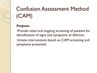
Objectives Definition Pathogenesis Causes ( neurological / non neurological) Risk factors. Clinical features. Diagnosis and management
Definition Disturbance in attention (reduced ability to focus, sustain, and shift attention) and awareness. The disturbance develops over a short period of time (usually hours to days). Represents a change from baseline. Tends to fluctuate during the course of the day. Additional disturbance in cognition (memory deficit, disorientation, language, visuospatial ability)
The disturbances are not better explained by another preexisting, evolving or established neurocognitive disorder. There is evidence from the history, physical examination, or laboratory findings that the disturbance is caused by a medical condition, substance intoxication or withdrawal, or medication side effect.
Additional features: Psychomotor behavioral disturbances such as hypoactivity, hyperactivity with increased sympathetic activity, and impairment in sleep duration. Variable emotional disturbances, including fear, depression, euphoria
Pathogenesis and localization lesions involving the ascending reticular activating system (ARAS) from the mid-pontine tegmentum rostrally to the anterior cingulate regions. Nondominant" parietal and frontal lobes.
Risk factors Polypharmacy (particularly psychoactive drugs) Infection. Dehydration. Immobility (including restraint use). malnutrition. Use of bladder catheters. Drugs.
Clinical presentation Disturbance of consciousness: One of the earliest manifestations of delirium is a change in the level of awareness and the ability to focus, sustain, or shift attention.. Distractibility,: one of the hallmarks of delirium, is often evident in conversation
Clinical presentation Change in cognition: Delirious individuals have cognitive and perceptual problems, including memory loss, disorientation, and difficulty with language and speech. Formal mental status testing can be used to document the degree of impairment. Ascertain the patient's level of functioning prior to the onset of delirium from family members, caregivers, or other reliable informants
Perceptual disturbances : . Hallucinations can be visual, auditory or somatosensory, usually with lack of insight. Hallucinations can be simple, e.g., shadows or shapes, or complex, as people and faces. Sounds can also consist of simple sounds or hearing voices with clear speech. Language difficulties: Patients may lose the ability to write or to speak a second language
Clinical presentation Prodromal features: Complaints of fatigue. Sleep disturbance (excessive daytime somnolence or insomnia). Depression. Anxiety, restlessness, irritability Hypersensitivity to light or sound.
Other features : psychomotor agitation. sleep-wake reversals Hypoactive delirium
EVALUATION There are two important aspects to the diagnostic evaluation of delirium: Recognizing that the disorder is present. Search for the underlying medical illness that has caused delirium.
History: recent febrile illness, history of organ failure medication list, history of alcoholism or drug abuse, or recent depression.
General examination: is often difficult or impossible in the confused or uncooperative patient. perform a focused assessment, concentrating upon vital signs, the state of hydration, skin condition, and potential infectious foci. The patient's general appearance may be suggestive, eg,, the jaundiced appearance of hepatic failure, the stigmata of renal failure. Needle tracks strongly suggest drug abuse. Cherry-red lips indicate possible carbon monoxide poisoning. The breath may smell of alcohol, fetor hepaticus, uremic fetor or ketones.
A bitten tongue or posterior fracture-dislocation of the shoulder suggests a convulsive seizure . Signs of head injury. Subhyaloid or retinal hemorrhages raise the possibility of an intracranial hemorrhage
Alcohol or sedative-drug withdrawal may cause a delirium characterized by autonomic nervous system activation (tachycardia, sweating, flushing, dilated pupils). Anticholinergic toxicity in elders can cause delirium without peripheral signs (eg, fever, mydriasis, tachycardia). Sepsis may present as delirium without obvious fever (sometimes even with hypothermia)
The physical signs of metabolic/toxic delirium can include nonrhythmic muscle jerking (multifocal myoclonus), flapping motions of an outstretched, dorsiflexed hand (asterixis), and postural action tremor.
Pitfalls in the examination: Temperature may be under 38.3 C even in the presence of serious infections. Auscultatory and radiographic findings of pneumonia may be subtle or absent abdominal serious conditions may present without peritoneal signs . False-positive findings occur as well (eg, nuchal rigidity may not signify meningitis).
Neurologic examination : Assessment of the level of consciousness, degree of attention or inattention, Visual fields, cranial nerve and motor deficit. Certain aspects of the examination may be difficult or unreliable in uncooperative patients (eg, sensory testing) Posterior cortical strokes, for example, can present as delirium with few findings other than hemianopia, and in some cases may present with no focal symptoms or signs.
The absence of focal examination findings does not exclude the possibility of focal or multifocal neurologic lesions as the cause of the delirium.
non neurological causes Fluid and electrolyte disturbances (dehydration, hyponatremia and hypernatremia) Infections (urinary tract, respiratory tract, skin and soft tissue) Drug or alcohol toxicity Withdrawal from alcohol Withdrawal from barbiturates, benzodiazepines, and selective serotonin reuptake inhibitors Metabolic disorders (hypoglycemia, hypercalcemia, uremia, liver failure, thyrotoxicosis) Low perfusion states (shock, heart failure) Postoperative states, especially in the elderly
Neurological causes Some neurological diseases can be misdiagnosed as confusion: Temporal-parietal : Patients with Wernicke's aphasia may appear delirious in that they do not comprehend or obey and seem confused. The problem is restricted to language, Other aspects of mental function are intact. Transient global amnesia (TGA): transient bitemporal dysfunction resulted in deficit restricted to memory lasting less 24 hrs.
Occipital: Anton's syndrome of cortical blindness and confabulation . Careful examination- visual loss. Frontal: Patients with bifrontal lesions (eg, from tumor or trauma) often show akinetic mutism, lack of spontaneity, lack of judgment, problems with recent or working memory, blunted or labile emotional responses, and incontinence.
Confusion or delirium due to acute or subacute brain lesions, such as stroke or multifocal white matter inflammation: may occur without focal deficits on examination.
Nonconvulsive status epilepticus : Under recognized. Requires an EEG for detection and continuous EEG for management. Often patients show no classic ictal features. Features suggesting the possibility of seizures: prominent bilateral facial twitching, unexplained sustained gaze during obtunded periods, automatisms (lip smacking, chewing, or swallowing movements), and acute aphasia or neglect without a structural lesion.
Dementia : May sometimes be confused with delirium Characteristic differences in progression and cognitive features usually distinguish these disorders. Cognitive change in Alzheimer disease is typically insidious, progressive, without much fluctuation, and occurs over a much longer time (months to years). Attention is relatively intact, as are remote memories in the earlier stages. Dementia with Lewy bodies (DLB) is similar to Alzheimer disease but can be more easily confused with delirium, because fluctuations and visual hallucinations are common and prominent.
Primary psychiatric illnesses: Delirium is commonly misdiagnosed as depression. Both are associated with poor sleep and difficulty with attention or concentration. Agitated depression may be especially problematic. depression is less fluctuating than delirium. Mania can be confused with hyperactive delirium with agitation, delusions, and psychotic behavior. mania is usually associated with a history of previous episodes of mania or depression. Schizophrenia the delusions are usually highly systematized, the history is longer, and the sensorium is otherwise clear.
Investigations Laboratory tests: Serum electrolytes, creatinine, glucose, calcium, complete blood count, and urinalysis and urine culture . Drug levels should be ordered where appropriate (digoxin, lithium, or quinidine). Toxic screen of blood and urine
Investigations Blood gas (ABG): respiratory alkalosis due to early sepsis, hepatic failure, early salicylate intoxication, or cardiopulmonary causes metabolic acidosis uremia, diabetic ketoacidosis, lactic acidosis, late phases of sepsis or salicylate intoxication, or toxins including methanol and ethylene glycol. Chest x-ray . Liver function tests. Thyroid function. Vitamin B12 levels.
Investigations Neuroimaging: Brain CT Neuroimaging may not be necessary if : the initial clinical evaluation discloses an obvious treatable medical illness , there is no evidence of trauma, no new focal neurologic signs are present.
Neuroimaging may still be required if : The delirium does not improve despite appropriate treatment of the underlying medical problem. Neurologic examination is confounded by diminished patient responsiveness or cooperation.
MRI is more sensitive than head CT for some conditions like acute stroke, posterior fossa lesions, and white matter lesions. It should be considered if brain CT is normal and there is still high suspicion of neurological etiology.
Lumbar puncture : Suspected CNS infections If the cause of delirium is not obvious. In febrile patients with delirium, even when alternate explanatory conditions for delirium are present or suspected. Neuroimaging should be obtained prior to lumbar puncture in patients with coma, focal signs, papilledema, or suspicion of increased intracranial pressure.
EEG: Triphasic waves are associated with hepatic encephalopathy or other severe metabolic disturbances including uremic and septic encephalopathy. Herpes simplex encephalitis may be associated with high amplitude periodic complexes in the temporal lobe leads.
Summary Delirium is a clinical syndrome caused by a medical condition, substance intoxication or withdrawal, or medication side effect that is characterized by a disturbance of consciousness with reduced ability to focus, sustain, or shift attention Nearly 30% of older medical patients experience delirium at some time during hospitalization. The incidence is higher in those with advanced age and pre-existing brain disease A disturbance of consciousness and altered cognition are essential components of delirium. Some patients are drowsy and lethargic, others are agitated and confused. Visual hallucinations, tremulousness, and myoclonus/asterixis are variably present
Focal or lateralized neurologic findings are not characteristic of delirium. A careful neurologic examination can also distinguish between focal syndromes that can mimic delirium The past medical history, a review of medications, and a physical examination may provide clues as to the underlying etiology Laboratory evaluation in patients with delirium should include serum electrolytes, creatinine, glucose, calcium, complete blood count, and urinalysis and urine culture. Drug levels, toxicology screen, liver function testing, and arterial blood gas should follow if the cause remains obscure Neuroimaging, lumbar puncture, and electroencephalogram are not required in most patients with delirium, but are recommended in specific clinical scenarios, including in those whose cause remains obscure after routine testing


