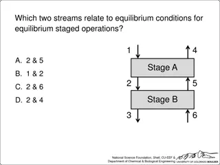
Understanding the Vertebral Column and Abnormal Curvatures
The vertebral column, extending from the cranium to the coccyx, plays vital roles in protecting the spinal cord, providing support, and facilitating movement. It consists of 33 vertebrae arranged in 5 regions, each with specific functions and characteristics. The column's curvatures, including cervical, thoracic, lumbar, and sacral, contribute to flexibility and shock absorption. Abnormal curvatures can arise from developmental anomalies or pathological processes, leading to conditions like kyphosis, lordosis, and scoliosis. Understanding the structure and functions of vertebrae is crucial for maintaining spinal health.

Download Presentation

Please find below an Image/Link to download the presentation.
The content on the website is provided AS IS for your information and personal use only. It may not be sold, licensed, or shared on other websites without obtaining consent from the author. Download presentation by click this link. If you encounter any issues during the download, it is possible that the publisher has removed the file from their server.
E N D
Presentation Transcript
Vertebral Column (Regional Characteristics of Vertebrae: not included) Ali Jassim Alhashli 20121098 Year IV Problem VII Musculoskeletal
Vertebral Column Extendingfrom cranium to the apex of coccyx. Functions: Protects the spinal cord and spinal nerves. Supports the weight of the body superior to the level of the pelvis. Provides partly rigid and flexible axis for the body. Pivot for the head. Plays an important role in posture and movement. It consists of 33 vertebrae arranged in 5 regions: 7 cervical. 12 thoracic. 5 lumbar. 5 sacral: fused in adults to for the sacrum. 4 coccygeal: fused to form the coccyx. Successive vertebrae bear increasing amounts of body s weight. The vertebrae reach maximum size immediately superior to the sacrum, which transfers the weight to the pelvic girdle at the sacroiliac joints. The vertebral bodies contribute of the height of the presacral vertebral column, and the fibrocartilage of IV discs contribute .
Curvature of Vertebral Column 4 curvatures: Cervical Thoracic Lumbar Sacral Function: provide a flexible support (shock- absorbing resilience) for the body. Thoracic and sacral curvatures which are (kyphoses). Cervical and lumbar curvatures: secondary curvatures which are (lordoses). Cervical curvature becomes prominent: when an infant begins to hold his/her head erect. Lumbar curvature becomes apparent: when an infant begins to walk and assumes the upright posture. The sacral curvature of females is reduced so that the coccyx protrudes less into the pelvic outlet (birth canal). curvatures: concave primary anteriorly convex anteriorly
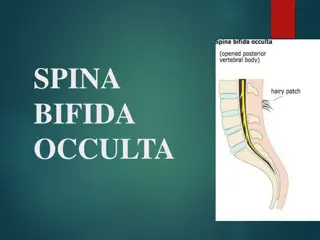
Abnormal Curvatures of Vertebral Column They result from: Developmental anomalies. Pathological processes (such as osteoporosis: vertebral body osteoporosis is more common in thoracic vertebrae). Excessive thoracic kyphosis: abnormal increase in the thoracic curvature. There will be overall loss of height. Note that kyphosis occurs in geriatric people of both sexes (normally). Excessive lumbar lordosis: abnormal increase in the lumbar curvature. Note that women develop a temporary lordosis during late pregnancy. Scoliosis (curved back): abnormal lateral curvature (S-shaped). Most common deformity of the vertebral column in pubertal girls (aged 12-15 years).
Structure and Function of Vertebrae A typical vertebra consists of: Vertebral body: Massive part of the vertebra. Gives strength and supports weight. Superior and inferior surfaces are covered with hyaline cartilage except at the periphery where there is a ring of smooth bone (epiphyseal rim). Vertebral arch: Lies posterior to the vertebral body. Formed by: right and left pedicles and laminae. Pedicles are short, thick processes which join the vertebral arch to the vertebral body. Laminae unite in the midline. Vertebral arch and posterior surface of vertebral body form the walls of the vertebral foramen. Vertebral notches: the superior and inferior vertebral notches of adjacent vertebrae contribute to the formation of the IV foramina, which give passage to spinal nerve roots and accompanying vessels and contain the spinal ganglia. Seven processes: One median spinous process: projects posteriorly (and usually inferiorly). Two transverse processes: project posterolaterally. Four articular processes: two superior and two inferior. They form zygapophyseal joints. Note: spinous process and the two transverse processes afford attachment for deep back muscles and serve as levers that help muscles move the vertebrae.
Joints of Vertebral Column Joints of vertebral bodies: Symphyses (secondary cartilaginous joints) designed for weight-bearing and strength. The articulating surfaces of adjacent vertebrae are connected by IV discs and ligaments. The IV discs provide strong attachments between the vertebral bodies permitting movment between adjacent vertebrae. Each IV disc consists of: Annulus fibrosus (outer fibrous part): ring, concentric lamellae of fibrocartilage which insert into the smooth rounded epiphyseal rim of vertebral bodies. Nucleus pulposus (gelatinous central mass): Central core, becomes broader when compressed and thinner when stretched. With age, the nuclei pulposi dehydrate and lose elastic and proteoglycans while gaining collagen, eventually becoming dry, granular and more resistant to deformation. Because the lamellae are thinner and less numerous posteriorly than they are anteriorly or laterally, the nucleus pulposus is not centered in the disc but is more posteriorly placed. Note that the nucleus pulposus is avascular. There is no IV disc between C1 (atlas) and C2 (axis) vertebrae. The most inferior functional disc is between L5 and S1 vertebrae. Thicker in cervical and lumbar regions and thinner in the superior thoracic region.
Joints of Vertebral Column Joints of vertebral bodies: Uncovertebral joints: located between the uncus of the bodies of the C3-C6 vertebrae. The articulating surfaces of these joint-like structures are covered with cartilage and contain a capsule filled with fluid. They are considered to be synovial joints. Anterior longitudinal ligament: Strong, broad, fibrous band. Covers and connects the anterolateral aspects of the vertebral bodies and IV discs. Extends from the pelvic surface of the sacrum to the anterior tubercle of the C1 vertebra (atlas) and occipital bone. Limits extension of the vertebral column. Posterior longitudinal ligament: Narrower, weaker and runs within the vertebral canal along the posterior aspect of the vertebral bodies. Prevents hyperflexion of the vertebral column and posterior herniation of the IV discs.
Joints of Vertebral Column Joints of vertebral arches: Zygapophyseal joints (facet joints): Synovial. Plane type. Between the superior and the inferior articular processes of adjacent vertebrae. Covered with joint (articular) capsule. Permit gliding movements between the articular processes. Accessory ligaments of intervertebral joints: The laminae of adjacent vertebral arches are joined by broad, pale, yellow elastic fibrous tissue called the ligamenta flava which extend almost vertically from the lamina above to the lamina below. Adjacent spinous processes are united by weak, almost membranous ligaments and strong fibrous supraspinous ligaments. interspinous
Joints of Vertebral Column Craniovertebral joints: they are synovial joints with no IV discs Atlanto-occipital joints: Between the lateral masses of C1 (atlas) and the occipital condyles. They allow flexion and extension (+bending and rotation). They are synovial joints of the condyloid type. Anterior and posterior atlanto-occipital membranes. Atlanto-axial joints (allowing rotation movement): Two (right and left) lateral atlanto-axial joints: plane-type between the lateral masses of C1 and the superior facets of C2. One median atlanto-axial joint: pivot joint. During rotation of the head, the dens of C2 is the pivot, which is held in a socket formed anteriorly by the anterior arch of the atlas and posteriorly by the transverse ligament of the atlas.
Herniation of Nucleus Pulposus Herniation or protrusion of the gleatinous nucleus pulposus into or through the anulus fibrosus is a well- recognized cause of low-back and lower limb pain. If degeneration of the posterior longitudinal ligament and wearing of the anulus fibrosus has occurred, the nucleus pulposus may herniate into the vertebral canal and compress the spinal cord or nerve roots of spinal nerves in the cauda equina. Herniations usually occur posterolaterally, where the anulus is relatively thin and does not receive support from the posterior or anterior longitudinal ligaments. Posterolateral herniation is most common in the lumbar region at the L4-L5 or L5-S1 levels. Sciatica: pain in the lower back and hip and radiating down the back of the thigh into the leg, is often caused by a herniated lumbar IV disc or osteophytes that compress the L5 or S1 component of the sciatic nerve.