Embryology: Stages of Development and Gestation Period in Different Animals
Embryology covers the Germinal, Embryonic, and Fetal stages of development in animals, highlighting processes like cleavage and formation of morula. Additionally, it provides insights into the gestation periods of various animals ranging from cows and mares to humans and elephants, each with its unique duration. The content also explores the migration patterns of zygotes and the variety in cleavage types seen across species.

Download Presentation

Please find below an Image/Link to download the presentation.
The content on the website is provided AS IS for your information and personal use only. It may not be sold, licensed, or shared on other websites without obtaining consent from the author. Download presentation by click this link. If you encounter any issues during the download, it is possible that the publisher has removed the file from their server.
E N D
Presentation Transcript
UNIT-8 EMBRYOLOGY Topic - General embryology (Periods of Embryology) Date of Lecture 07.04.2014 Course instructor Dr. Manoj Kumar Sinha Department of veterinary Anatomy
PERIODS OF EMBRYOLOGY The changes occur during the whole gestation period are divided into three major stages: Germinal Stage : Includes Cleavage, Formation of morula and blastocyst, implantation of blastocyst, and appearance of three germinal layer Embrryonic Stage: Includes differentiation of three germinal layers and and formation of most of the tissues and organs Foetal Stage: Includes growth of various system of embryo without much tissue differentiation and development of placenta
GESTATION PERIOD OF DIFFERENT ANIMALS ANIMALS PERIOD OF GESTATION (IN DAYS) Cow 280-290 Mare 330-340 Dog/Cat 60-65 Sow 110-130 Goat/Sheep 140-160 Human 270-290 Elephant 600-650 Rabbit 30-35 Rhinoceros 520-560 Rat 20-25 Monkey 160-175 Hen 20-25 (Incubation Period)
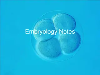
GERMINAL STAGE CLEAVAGE : Is the process of rapid successive mitotic division of an ovum immediately after its fertilization ,resulting in production of increasing number of smaller cells within the zona pellucida ,called BLASTOMERS The rate of cleavage division = 1/Amount of yolk present in ova TYPES OF CLEAVAGE: Vary species to species Holoblastic/Total cleavage (scanty deutoplasm in ovum) (Abundant deutoplasm in ovum) Meroblastic/Partial cleavage Equal Eg: Mammals Unequal Eg: Amphibians Eg: Reptiles Discoidal Suprrficial Eg: Arthropod
MORULA No. of cells (Blastomeres) 12 to 16 and remain closely packed without formation of any cavity looks like a mulberry fruit, called Morula In chick embryo it appears as a disc shaped mass of cells Central cell mass of the morula gives rise embryo proper and peripheral cells from protective and nutritive covering of the embryo With the continuation of cleavage the zygote migrates from ampulla of fallopian tube and reach to the cavity of uterus either in the horn or in the body depends upon the species Migration depends upon cilia of the mucous membrane of the tube and muscular contraction
BLASTOCYST Blastocyst formed b/w 4-5 day after fertilization in cow and man In the uterine cavity the blstomers continue to divide ,fluid from the lumen of the uterus enter inside morula through zona pellucida and form central cavity This fluid separates blstomeres into an inner cell mass and an outer cell mass This whole structre are called blastocyst and fluid filled cavity is called Blastocele Outer cell mass which form the wall of blastocyst is called Trophoblst and iner cell mass is called Embryoblast The trophoblast that covers the embryonic pole of of blastocyst is k/a Polar Trophoblast and other part forming the wall of cavity is known as Mural Trophoblast
IMPLANTATION OF BLASTOCYST The zona pellucida disappears on 5thor 6thday of fertilization in human and 7thto 9thday in cow By the end of 2ndwk the blastocyst is completely embeded in the endometrial stoma in human by histolytic action of trophoblast (Invasive process) In domestic animal it is not invasive process (except in Pig) but it occures by adesion and apposition between trophoblast and uterine epithelium . In Ruminants pacental attachments involves both caruncular and inter-caruncular area of endometrium On 17thday of gestation uninucleate trophoblst give rise to binucleate cells it persist throughout gestation period and give rise to immunoloic protection In Mare implantation takes place on 24-40 days Trophoblast differentiate into inner (cytotrophoblast) outer (syncytotrophoblast/syncytium)
Continue... The embryoblast give rise to an inner endodermic layer thus bilamilar blastocyst is formed. The endodermic layer of embryoblast is k/a Hypoblast, the cells close to polar trophoblast of embryoblast are transformed into columnar cell k/a Epiblast(Primary Ectoderm) The primary endoderm enclose a cavity opposite to embryonic pole k/a Primary yolk Sac The cytotrophoblast and epiblast are separated with fluid filled cavity k/a Primary Amniotic Cavity Therefore a bilaminar plate (bilaminar germ disc) formed between primary yolk sac and primary amniotic cavity which gives rise to the development of embryo
Appearance of Germ Layers At first germ disc is oval and subsequently it becomes elongated in shape having broad cephalic end and other narrow caudal end Ectodermal layer of germ disc differentiated into 3 functional zone 1) Surface ectoderm It give rise to epidermis of skin 2) Neural plate It gives rise to future nervous system 3) Pluripotent cellular zone Fast proliferating cells and form linear opacity in the midline called Primitive Streak(Pluripotent cell) Pluripotent cells migrate and invaginate into the space between epiblast and hypoblast k/a Gastrulation Formation of three germ layer is necessary for organogenesis i. Endoderm formed the epiblast by invading through hypoblast ii. Mesoderm cells of epiblast migrate bilaterally through primitive streak reach b/w epiblast and endoderm to form mesoderm iii. Ectoderm Epiblast is called ectoderm
Formation of Notochord The cephalic end of the primitive streak becomes swollen called Primitive Knot or Henson s node A central depression appears at henson s node called Primitive pit (Blastopore) Gradually primitive pit extend cranially and at the same time cavity of primitive pit also extend and converts Notochordal canal Gradually notochordal wall becomes flat to form a notochordal plate and this plate forms a fold which becomes deep to form a tube and converted into solid cord of cells called Notochord Notochord acts as forerunner for development of vertebral column Later on notochord disappeared and parts of it persist as nucleus pulposus of invertibral disc it into a canal called