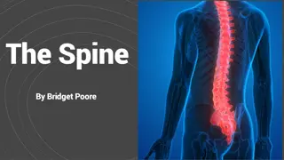
Understanding Spinal Canal Stenosis: Causes, Symptoms, and Classification

SPINAL CANAL STENOSIS
Spinal canal stenosis
STENOSIS- Constriction of a tube.
Spinal canal stenosis is defined as an abnormal
narrowing of
Osteoligamentous
vertebral canal
/ or the
intervertebral foramina
, causing direct compression or
compromise of the dural sac / caudal nerve root or their
vasculature. Producing symptom of rediculopathy or
claudication.
Refers to narrowing of the spinal canal, nerve root
canals or intervertebral foramina due to spondylosis
& degenerative disc disease.
Usually occurs in
cervical
&
lumbar spine.
CERVICAL STENOSIS
Usually present with cervical radiculopathy: radiating arm pain
with numbness and paresthesia and occasionally, associated
weakness.
If the stenosis is severe enough or if it is
positioned centrally in the spine, patients may
present with signs and symptoms of myelopathy (
Spinal cord dysfunction): Finger numbness,
clumsiness and
difficulty walking due to spasticity and loss of
position sense.
In more severe cases, the patient can have bowel
and bladder control dysfunction. Upon
examination, these patient have ‘long tract signs’
such as hyper-reflexia and clonus.
Lumbar stenosis
The further narrowing of the
anatomically narrower spinal canal
in the lower lumbar region due to a
structural abnormality is termed as
Spinal stenosis. Since this occurs
most commonly in the lumbar spine,
it is known as
Lumbar stenosis.
It present as backache with
radicular pain in the legs, brought
on by exercise or walking for a
perticular distance
CLASSIFICSATION-
1)
Congenital or developmental stenosis-
The
lower lumbar canal become narrow due to
laminar thickening of the developmental origin
or congenital as seen in achondroplastic
dwarfs.
2)
Acquired stenosis-
The canal become
narrow over a period of time due to various
causes-
a)
Degenerative disease-
Osteophytes
encroach upon the space of the lumbar
canal & cause compression.
b)
Degenerative spondylolisthesis-
The canal become narrow due
to slip of the vertebrae, perticularly at L4-L5 level.
c)
Miscellaneous-
conditions such as fluorosis and paget’s disease are
associated with narrow lumbar canal.
CLINICAL FEATURES-
The patients classically present with symptoms of
intermittent claudication. He develops pain heaviness or
parasthesia in the lower limb after walking for a certain
distance.
The symptom become so severe that he has to take rest
immediately for a few minutes. The symptoms settle down
temporarily & allow him to walk further the same distance.
NEUROLOGICAL EXAMINATION-
Neurological examination of the lower limb
may be normal or may be bizarre
neurological deficit.
In severe cases, there may be signs of cauda
equina compression with loss of bladder &
bowel control.
INVESTIGATIONS-
1)
Plain radiograph of the spine- Measurement
of the lumbar canal will show decreased
anteroposterior diameter of the canal or
narrowing of the interlaminar space.
2)
Myelography- is useful in the diagnosis . It
shows multiple indentations in the dye
column.
3)
CT Scan & MRI are also helpful in the
diagnosis.
TREATMENT-
The treatment could be-
1)
Conservative
2)
Surgical
1)
Conservative treatment-
The conservative treatment
approach is basically to emphasize flexion exercises &
generalized flexion attitudes, avoiding extension.
Drug to control pain & inflammation.
Improve strength endurance & tone of the
abdominal muscle-
This greatly helps in the
maintenance of the flexed posture, which
provides comfort to these patients.
Back ergonomics, avoiding extension
attitudes are taught.
Lumbar traction may be beneficial if
comfortable.
Lumbar corset should be used during
strenuous activities.
Gentle passive manipulation technique is
effective.
SURGICAL TREATMENT-
The aim of surgery is to decompress the cord
by performing laminectomy.
THANK YOU
Spinal canal stenosis is the abnormal narrowing of the spinal canal or intervertebral foramina, leading to compression of nerves and blood vessels. It can result in symptoms such as radiculopathy, claudication, myelopathy, and more. Cervical stenosis presents with arm pain and weakness, while lumbar stenosis causes backache and leg pain. Classification includes congenital and acquired causes, with degenerative disease being a common factor.

Download Presentation

Please find below an Image/Link to download the presentation.
The content on the website is provided AS IS for your information and personal use only. It may not be sold, licensed, or shared on other websites without obtaining consent from the author. Download presentation by click this link. If you encounter any issues during the download, it is possible that the publisher has removed the file from their server.
E N D
Presentation Transcript
Spinal canal stenosis STENOSIS- Constriction of a tube. Spinal canal stenosis is defined as an abnormal narrowing of Osteoligamentous vertebral canal / or the intervertebral foramina, causing direct compression or compromise of the dural sac / caudal nerve root or their vasculature. Producing symptom of rediculopathy or claudication. Refers to narrowing of the spinal canal, nerve root canals or intervertebral foramina due to spondylosis & degenerative disc disease. Usually occurs in cervical & lumbar spine.
CERVICAL STENOSIS Usually present with cervical radiculopathy: radiating arm pain with numbness and paresthesia and occasionally, associated weakness.
If the stenosis is severe enough or if it is positioned centrally in the spine, patients may present with signs and symptoms of myelopathy ( Spinal cord dysfunction): Finger numbness, clumsiness and difficulty walking due to spasticity and loss of position sense. In more severe cases, the patient can have bowel and bladder control dysfunction. Upon examination, these patient have long tract signs such as hyper-reflexia and clonus.
Lumbar stenosis The further narrowing of the anatomically narrower spinal canal in the lower lumbar region due to a structural abnormality is termed as Spinal stenosis. Since this occurs most commonly in the lumbar spine, it is known as Lumbar stenosis. It present as backache with radicular pain in the legs, brought on by exercise or walking for a perticular distance
CLASSIFICSATION- 1) Congenital or developmental stenosis- The lower lumbar canal become narrow due to laminar thickening of the developmental origin or congenital as seen in achondroplastic dwarfs. 2) Acquired stenosis- The canal become narrow over a period of time due to various causes- a) Degenerative disease- Osteophytes encroach upon the space of the lumbar canal & cause compression.
Degenerative spondylolisthesis- The canal become narrow due to slip of the vertebrae, perticularly at L4-L5 level. b) Miscellaneous- conditions such as fluorosis and paget s disease are associated with narrow lumbar canal. CLINICAL FEATURES- c) The patients classically present with symptoms of intermittent claudication. He develops pain heaviness or parasthesia in the lower limb after walking for a certain distance. The symptom become so severe that he has to take rest immediately for a few minutes. The symptoms settle down temporarily & allow him to walk further the same distance.
NEUROLOGICAL EXAMINATION- Neurological examination of the lower limb may be normal or may be bizarre neurological deficit. In severe cases, there may be signs of cauda equina compression with loss of bladder & bowel control. INVESTIGATIONS- 1) Plain radiograph of the spine- Measurement of the lumbar canal will show decreased anteroposterior diameter of the canal or narrowing of the interlaminar space.
2) Myelography- is useful in the diagnosis . It shows multiple indentations in the dye column. 3) CT Scan & MRI are also helpful in the diagnosis. TREATMENT- The treatment could be- 1) Conservative 2) Surgical 1) Conservative treatment- The conservative treatment approach is basically to emphasize flexion exercises & generalized flexion attitudes, avoiding extension.
Drug to control pain & inflammation. Improve strength endurance & tone of the abdominal muscle- This greatly helps in the maintenance of the flexed posture, which provides comfort to these patients. Back ergonomics, avoiding extension attitudes are taught. Lumbar traction may be beneficial if comfortable. Lumbar corset should be used during strenuous activities. Gentle passive manipulation technique is effective.
SURGICAL TREATMENT- The aim of surgery is to decompress the cord by performing laminectomy.


 Myelography- is useful in the diagnosis . It")
 Myelography- is useful in the diagnosis . It")