Embryology of Spinal Cord and Vertebral Column Development
Explore the fascinating embryological journey of the spinal cord and vertebral column development, covering topics such as neural tube formation, layers of the spinal cord, subdivisions of mantle and marginal zones, chondrification, ossification stages, spina bifida types, and more. Dive into the stages of neural tube development and understand the critical processes involved in shaping these essential structures during early human development.

Download Presentation

Please find below an Image/Link to download the presentation.
The content on the website is provided AS IS for your information and personal use only. It may not be sold, licensed, or shared on other websites without obtaining consent from the author. Download presentation by click this link. If you encounter any issues during the download, it is possible that the publisher has removed the file from their server.
E N D
Presentation Transcript
Development of Spinal Cord Development of Spinal Cord & Vertebral Column & Vertebral Column Neuroanatomy block-Embryology -Lecture 1 Editing file
Color guide Color guide: : Only in boys slides in Green Only in girls slides in Purple important in Red Red Notes in Grey Grey Green Purple Objectives Objectives At the end of the lecture, students should be able to: 1. 1. 2. 2. 3. 3. 4. 4. 5. 5. Describe the development of the spinal cord from the neural tube Describe the development of the spinal cord from the neural tube. . List the layers of the spinal cord and its contents List the layers of the spinal cord and its contents. . List subdivisions of mantle & marginal zones List subdivisions of mantle & marginal zones List meningeal layers and describe positional changes of spinal cord List meningeal layers and describe positional changes of spinal cord. . Describe development of vertebral column from sclerotomic portion of Describe development of vertebral column from sclerotomic portion of paraxial mesoderm paraxial mesoderm. . Describe chondrification & ossification stages in vertebral development Describe chondrification & ossification stages in vertebral development. . Describe spina bifida and its types Describe spina bifida and its types 6. 6. 7. 7.
Introduction Introduction Found only in girl s slides It has 4 components : Third week Third week 1 - Hypoblast 3 - Epiblast It forms Three Germ Layers 1) 1) Ecto Ectoderm 2) 2) Meso Mesoderm 3) 3) Endo Endoderm It has also 2 - Yolk sac cavity 4 - Amniotic cavity Second week Second week 1) 2) Primitive node Primitive streak Development of Neural Tube Development of Neural Tube The Neural Tube is a derivative of the ectoderm Gives rise to the brain and the spinal cord It has 1 ) Cervical flexure Cephalic flexure 2 ) 3 3
Development of Neural Tube Development of Neural Tube Stages of development: 1 4 2 3 The margins of the neural plate rehto hcae ot hcaorppa eht mrof ot esuf dnaneural tube The neural plate folds to form longitudinal groove )evoorg laruen( )evoorg laruen(with prominent neural folds neural folds. Ectodermal cells dorsal to notochord thicken to form the neural plate neural plate. Notochordstimulates neural tube formation which in turn stimulates development of the vertebral column. )sdlof laruen( )sdlof laruen( neural tube. The tube then separates from the overlying ectoderm. Notochordinduces the ectoderm above it which thickens to form the neural plate. 5 Closer of the neural tube begins in the future neck region (4th somite) Then proceeds cranially and caudally The most cranial and caudal ends of the tubes still open as Anterior neuropores will close at day Posterior neuropores will close at day 27 The lumen of the tube will give ventricles of the brain and central canal of spinal cord. Somite 25 lamina terminalis 4 4
Development of the Spinal Cord Development of the Spinal Cord The spinal cord develops from the caudal 2/3of the neural tube The cells of the neural tube The cells of the neural tube are arranged in three layers alar plate ventricular zone marginal zone mantle zone marginal zone Inner undifferentiated cells Middle cell bodies of neurons Outer nerve fibers or axons of neurons (future white matter) ventricular zone sulcus limitans mantle zone ( future gray matter) Neurons of mantle layer differentiate into: basal plate ventral basal plate )nroh lartnev erutuf( snoruen rotom gniniatnoc dorsal alar plate ( future dorsal horn ) containing sensory neurons The 2areas are separated by a longitudinal groove (sulcus limitans(. 5 5
Mantle Layer of Spinal cord Mantle Layer of Spinal cord )rettam yarg erutuf( )rettam yarg erutuf( Proliferation and bulging of both alar& basalplates result in: 2 3 1 Narrowing of the lumen of the neural tube to form a small central canal central canal . Formation of dorsal median dorsal median septum septum . . Formation of ventral median ventral median fissure fissure. . Marginal Layer of Spinal cord Marginal Layer of Spinal cord )rettam etihw erutuf( )rettam etihw erutuf( dorsal dorsal funiculi funiculi increases in size due to addition of ascending, descending & intersegmental nerve fibers. it is divided into : dorsal dorsal ,lateral lateral dnaventral funiculi ventral funiculi )nmuloc etihw( Myelination of nerve fibers starts at 4th month & continues during the 1st postnatal year. Motor Motor erofeb detanileym srebifsensory sensory srebif . So, After a nerve injury, both motor and sensory axons have the ability to regenerate and, given a proper pathway. lateral lateral funiculi funiculi . ventral ventral funiculi funiculi the cells that are still in ventricular zone, became ependyma for the central canal. 6 6
Meninges Meninges These Are 3Membranes covering the neural tube: Outer thick dura mater Origin: Meso Mesodermal 1 1 Middle arachnoid mater origin: Ecto Ectodermal 2 2 subarachnoid subarachnoid ytivac a si :ecaps & dionhcara eht neewteb sraeppa htiw dellif semoceb retam aip eht )FSC( diulf lanipsorberec 3 3 Inner thin pia mater origin : Ecto Ectodermal 4 4 Positional Changes of Spinal Cord Positional Changes of Spinal Cord Initially, the spinal cord occupies the whole As a result a faster growth of vertebral column, the caudal end of spinal cord (conus medullaris conus medullaris level rehgih a ot yllaudarg stfihs ) . whole lanac larbetrev eht fo htgnel . The spinal cord in 3rd monthsame length as vertebral canal At birthspinal cord terminates at 3rd lumbar vertebra In adult spinal cord terminates at L1 8weeks 1. 2. 7 7
Specialization of Mesoderm Specialization of Mesoderm Appearance of the notochord (first sign) Three collections of the mesoderm appear lateral to the notochord Somites develop from the para-axial mesoderm. Intermediate mesoderm Double sheets of lateral plate mesoderm Intraembryonic Mesoderm Intraembryonic Mesoderm Located: between Ectoderm & Endoderm EXCEPTin the central axis of embryo where NOTOCHORD is found. Differentiates into Differentiates into 3 3 parts parts: : Dermatome Somite Paraxial mesoderm Myotome Divides into segments called somites . Sclerotome Intermediate mesoderm Lateral mesoderm 8 8
Development of the Vertebral Column Development of the Vertebral Column The vertebral column develops from the ventromedial parts (sclerotomes) of the somites Each one of somites divide into 3 parts: Sclerotome: form the vertebrae & ribs Dermatome: forms the dermis of the skin on the dorsal part of the body Myotome: forms the skeletal muscles of the neck, trunk & limbs Formation of Body of Vertebra At 4th week, each sclerotome becomes subdivided into two parts : a cranial part, consisting of loosely arranged cells a caudal part, of more condensed tissue. 1 cranial part The caudal part of each somite fuses with the cranial partof the consecutive somite,around the notochord to form the body of the vertebra, called the centrum. each centrum develops from 2adjacent sclerotomes. 2 centrum 3 the bodies of the vertebrae are intersegmental in origin. caudal part 9 9
Fate of Notochord In the region of the bodies of vertebrae: It degenerates. Between bodies of vertebrae: It forms the central part, nucleus pulposus of the intervertebral discs. Annulus fibrosus of the intervertebral discs is formed by the mesoderm surrounding the notochord. vertebral arch. Vertebral Development The fused sclerotomes grow dorsally around the neural tube and form the vertebral (neural) arch. Ventrolaterally, costal processes develop that give rise to ribs in thoracic region. The development occurs in 5 stages: All centers unite around 25 years ) Primary Ossification ( 3 centers) 3 ) Stage of Fusion (Fusion of 2V.arches) 4 ) Mesenchymal 1 ) Chondrification 2 ) Secondary Ossification 5 Fusion between halves of neural archoccursat 3 - 5 years, between neural arch & bodyat Appear atpuberty Appear at6th week Appear at the end of 8th week 3 - 6 years 10 10
Curvatures of Vertebral Column Curvatures of Vertebral Column Curvatures of Vertebral Column Curvatures of Vertebral Column Primary Secondary (convex anterior) (concave anterior) develop prenatally contains develop postnatally contains Thoracic Pelvic or Sacral Cervical Lumbar concave posteriorly as a result of lifting the head concave posteriorly as a result of walking Help support trunk, upper body 11 11
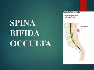
Spinal Cord Anomalies Spinal Cord Anomalies Spina Bifida Spina Bifida Failure of fusion of the halves of vertebral arches Incidence: 0.15% - 0.04 Sex: more frequent in females Has 2 types Occulta 20% Spinal dermal sinus Tethered cord Manifesta (cystic) 80% 1. Meningocele 2. Meningomyelocele 3. Myelochisis ) 2 ) 1 A. A. Spina Bifida Occulta Spina Bifida Occulta The closed type Only one vertebra is affected No clinical symptoms Skin overlying it is intact Sometimes covered by a tuft of hair Usually does not involve underlying neural tissue 12 12
B. Spina Bifida Cystica B. Spina Bifida Cystica Cystica is the most severe and complex form of spina bifida . It usually involves serious neurological problems . A portion of the nerves and the spinal cord are exposed outside the body Neurological symptoms are present It Subdivided into It Subdivided into: : . . Spina bifida with Spina bifida with meningomyelocoele meningomyelocoele 2 2 1 1 . . Spina bifida with meningocele Spina bifida with meningocele . . Spina bifida with myeloschisis Spina bifida with myeloschisis 3 3 protrusion of sac containing meninges with spinal cord and/or nerve roots. spinal cord is open due to failure of neural folds to develop. protrusion of sac containing meninges & cerebrospinal fluid. 13 13
Practice Practice Q Q A. Paraxial B. Intermediate C. Lateral D. Notochord Q Q ta trap laduac & lainarc otni dedividbus semoceb emotorelcs hcaE : A. 5th week B. 4th week C. 4th month D. 2nd month Q Q si nmuloc lanips fo erutavruc yradnoces ehT : A. Concave anterior B. Concave posterior C. Convex anterior D. Both B&C Q Q 8 8 : : regarding spinal bifida which one of the statement is correct ? A. The closed type is more frequent than the open B. The closed type presents with clinical symptoms C. In cases of spina bifida with meningocele, the spinal cord is open. D. Spina bifida is due to failure of fusion between the halves of vertebral arch. setimos dellac stnemges otni sedivid mredosem fo trap hcihW ? 5 5 : : Q Q 1 1 : : The neural tube is derivative from: A. endoderm B. mesoderm C. intermediate mesoderm D.ectoderm Q Q noruen rotom fo seidob llec sniatnoc droc lanips fo snoiger gniwollof eht fo eno hcihW ? A. Alar plate B. Basal plate C. Mantle zone D. Dorsal funiculus Q Q gnirud seunitnoc dna___ ta strats srebif evren fo noitanileyM ___________ . A. 2nd month -1st postnatal year B. 4th month -1st postnatal year C. 2nd month -2nd postnatal year D. 4th month -2nd postnatal year Q Q 4 4 : : morf detanigiro retam aruD : A. endoderm B. mesoderm C. intermediate mesoderm D.ectoderm 6 6 : : 2 2 : : 7 7 : : 3 3 : : 14 14 Answers:Q )D( 8 Q )D( 7 Q )B( 6 Q )A( 5 Q )B( 4 Q )B( 3 Q )B( 2 Q )D( 1
Members board Members board Team leaders Team leaders Abdulrahman Shadid Abdulrahman Shadid Ateen Almutairi Ateen Almutairi Girls team Girls team: : Boys team Boys team : : Ajeed Al Rashoud Ajeed Al Rashoud Taif Alotaibi Taif Alotaibi Noura Al Turki Noura Al Turki Amirah Al Amirah Al- -Zahrani Alhanouf Al Alhanouf Al- -haluli Sara Al Sara Al- -Abdulkarem Abdulkarem Renad Al Haqbani Renad Al Haqbani Nouf Al Humaidhi Nouf Al Humaidhi Jude Al Khalifah Jude Al Khalifah Nouf Al Hussaini Nouf Al Hussaini Rahaf Al Shabri Rahaf Al Shabri Danah Al Halees Danah Al Halees Rema Al Mutawa Rema Al Mutawa Amirah Al Dakhilallah Amirah Al Dakhilallah Maha Al Nahdi Maha Al Nahdi Razan Al zohaifi Razan Al zohaifi Ghalia Alnufaei Ghalia Alnufaei Mohammed Al Mohammed Al- -huqbani Salman Alagla Salman Alagla Ziyad Al Ziyad Al- -jofan jofan Ali Aldawood Ali Aldawood Khalid Nagshabandi Khalid Nagshabandi Omar Alammari Omar Alammari Sameh nuser Sameh nuser Abdullah Basamh Abdullah Basamh Alwaleed Alsaleh Alwaleed Alsaleh Mohaned Makkawi Mohaned Makkawi Abdullah Alghamdi Abdullah Alghamdi huqbani Zahrani haluli Contact us Contact us : : Editing file Editing file most probably you don't need this