Comprehensive Guide to Stomach Anatomy, Histology, and Barium Meal Radiology
Learn about the intricate anatomy of the stomach, its histological structure, lymphatic drainage, and the use of barium meal radiology for diagnostic purposes. Discover indications and contraindications, patient preparation, and clinical scenarios where these procedures are essential.

Download Presentation

Please find below an Image/Link to download the presentation.
The content on the website is provided AS IS for your information and personal use only. It may not be sold, licensed, or shared on other websites without obtaining consent from the author. Download presentation by click this link. If you encounter any issues during the download, it is possible that the publisher has removed the file from their server.
E N D
Presentation Transcript
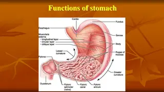
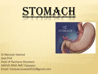
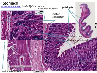
ANATOMY OF STOMACH SURFACES: Anterosuperior & Posteroinferior. BORDERS :lesser & greater curvatures. DIVIDED INTO:- Fundus, Body, Antrum & pylorus.
Short obese people : Transverse & Steer horn shape. Tall thin people: Longitudinal & J Shaped Max capacity :1500ml. Completely covered by peritoneum.
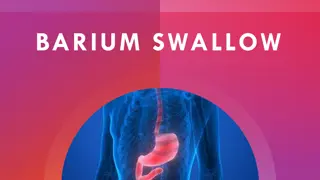
BARIUM MEAL Radiological study of stomach , duodenum, and proximal jejunum by oral administration of contrast media(barium sulphate).
Ba fills the lumen, taking the shape of the organ and delineates any abnormal structure. Radiolucencies are produced by mass lesions displacing Ba. Radio-opacities by ulcer, fistula, diverticulum which extends beyond normal contours of organ.
INDICATIONS Symptoms epigastric pain,anorexia,wt loss,dyspepsia,etc. Upper abdominal mass. GI Hemorrhage. Gastric or duodenal obstruction. Ca GEO junction,stomach,duodenum. Motility disorders. Systemic diseases viz-TB . CHILDREN-GE reflux, pyloric obstruction, malrotation.
CONTRAINDICATIONS Gastroduodenal perforation. H/o aspiration. Large bowel obstruction. Fistulous communication. Recent biopsy.
PREPARATION OF PATIENT NBM for 6hrs. Restrain smoking. In diabetes : early morning appt. In Gastric outlet obstruction prolonged fasting , metaclopramide given, sometimes nasogastric intubation & aspiration needed.
SINGLE CONTRAST STUDY Low density barium suspension (80- 100% w/v) is used. Conventional Radiography is done at 80-90kV. 10-15 ml of Ba given with patient supine & rotated in clockwise manner to obtain good mucosal coating. 100-250ml of Ba is given & films are taken in various views.
STANDARD VIEWS FOR SINGLE CONTRAST SUPINE FUNDUS ERECT OR PRONE BODY ANTRUM & PYLORUS PRONE RT SIDE DOWN D1 & C LOOP PRONE RT SIDE DOWN D4 SUPINE
Barium Sulphate 30% w/v is used. Radiography done at 120-130 kV. Permits visualization through the Barium column.
ADVANTAGES OF SINGLE CONTRAST Pylorospasm, fistulae & enlarged gastric rugae are better seen. Filling defects due to large masses in pyloric & duodenal region are more easily identifiable. Procedure of choice for suspected gastric or duodenal obstruction.
DISADVANTAGES Lack of sensitivity in detecting Small erosions/ulcers. Early gastric Ca. Subtle mucosal abnormalities.
DOUBLE CONTRAST STUDY High density (200-250% w/v) low viscosity Ba Sulphate, to coat the wall. Gas producing agents are used to distend the lumen, acts as negative contrast. Smooth muscle relaxant - Buscopan
STANDARD VIEWS FUNDUS PRONE RT SIDE DOWN BODY SUPINE WITH HEAD END ELEVATION ANTRUM & PYLORUS SUPINE RT SIDE UP D1 & C LOOP SUPINE RT SIDE UP PRONE RT SIDE DOWN D4
ADVANTAGES Highly accurate in detecting abnormalities following:- Gastric surgery Bile reflux gastritis Marginal ulcerations Recurrent Ca
DISADVANTAGES Probably misses some polyps, ulcers, erosions, especially in non dependent walls of stomach.
BIPHASIC STUDY 60-100% low viscosity Ba Sulphate. Mucosal relief film. 200-250 ml of Ba given in supine position with Eno in last few mouthfuls.
Prone Rt side down Duodenal cap & C loop in SC. Supine with Rt side up oblique Duodenum in DC. Erect film with compression Duodenal Bulb. Compression film & Fluoroscopy of stomach For contour defects, peristalsis & emptying.
Erect DC films of fundus in 2 views. Supine , 60o head up & DC film Upper body stomach. Supine , table horizontal & DC film lower body & pyloric antrum. Erect Rt Anterior Oblique for stomach, Duo & proximal Jejunum on 1 film.
AREAE GASTRICAE Micromucosal pattern consists of mosaic of tiny raised nodules(2- 4mm) with intervening grooves. Obvious in distal two third.
COMPLICATIONS 1. Leakage of Ba from an unsuspected perforation. 2. Aspiration pneumonia. 3. Barium impaction. 4. Acute gastric dilatation. 5. Barium embolisation if a bleeding ulcer is present.
THANK YOU