Overview of Esophagus and Stomach Imaging Techniques
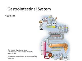
This content provides information on various imaging modalities used in examining the esophagus, including CT scan, MRI, Fluoroscopy, US, X-ray, and Nuclear medicine. Detailed images and descriptions of esophageal anatomy, abnormalities, and stomach anatomy are included, along with explanations of procedures like Fluoroscopy and CT scans for diagnostic purposes.

Download Presentation

Please find below an Image/Link to download the presentation.
The content on the website is provided AS IS for your information and personal use only. It may not be sold, licensed, or shared on other websites without obtaining consent from the author. Download presentation by click this link. If you encounter any issues during the download, it is possible that the publisher has removed the file from their server.
E N D
Presentation Transcript
RADIOLOGY OF ESOPHAGUS AND STOMACH
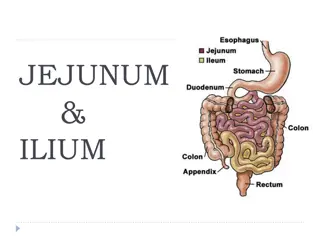
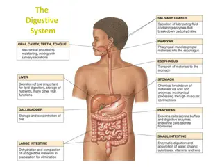
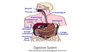
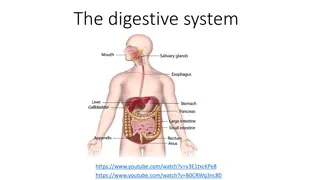
ESOPHAGUS ANATOMY
What are the modalities can be used in imaging the esophagus? A. CT scan B. MRI C. Fluoroscopy D. US E. X-ray F. Nuclear medicine
What are the modalities can be used in imaging the esophagus? A.CT scan B. MRI C. Fluoroscopy D. US E. X-ray F. Nuclear medicine
5 2 4 1 3 1
5 2 4 1 3 1 1. Esophagus 2. Trachea 3. Aorta 4. Lung 5. Pulmonary artery
Fluoroscope x- Real-time x ray. ray video Multiple sequential images Spot films
2 1 4 3
1-esophageal vestibule (ampulla). 2- B ring. 3- gastroesophageal junction. 4- stomach (gastric fundus). 2 1 4 3
Bird's beak appearance
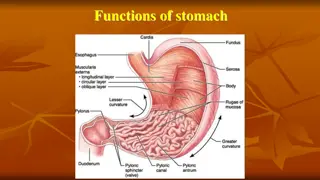
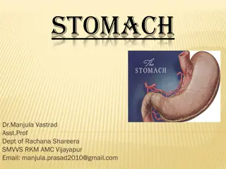
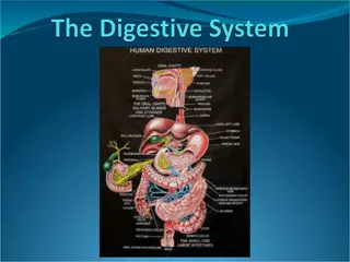
STOMACH ANATOMY
1 3 5 2 4 6
1 1. Stomach fundus 2. Greater curvature 3. Lesser curvature 4. Antrum 5. Pylorus 6. Mucosal folds 3 5 2 4 6
CT SCAN MRI
2 3 5 4 1
2 3 5 4 1 1-Greater curvature 2-Lesser curvature 3-Stomach fundus 4-Antrum 5-Pylorus
2 1 3 4 6 5
1. Stomach 2. Liver 3. Antrum 4. Pylorus 5. Spleen 6. Kidney 2 1 3 4 6 5


")